
Page 65 - Read Online
P. 65
Page 8 of 12 Hambright et al. Mini-invasive Surg 2024;8:19 https://dx.doi.org/10.20517/2574-1225.2024.08
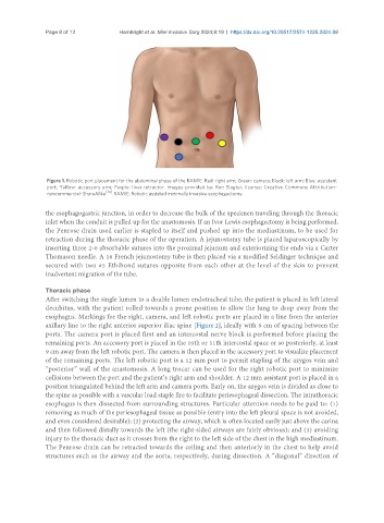
Figure 1. Robotic port placement for the abdominal phase of the RAMIE. Red: right arm; Green: camera; Black: left arm; Blue: assistant
port; Yellow: accessory arm; Purple: liver retractor. Images provided by: Ron Slagter, license: Creative Commons Attribution-
noncommercial-ShareAlike [34] . RAMIE: Robotic assisted minimally invasive esophagectomy.
the esophagogastric junction, in order to decrease the bulk of the specimen traveling through the thoracic
inlet when the conduit is pulled up for the anastomosis. If an Ivor Lewis esophagectomy is being performed,
the Penrose drain used earlier is stapled to itself and pushed up into the mediastinum, to be used for
retraction during the thoracic phase of the operation. A jejunostomy tube is placed laparoscopically by
inserting three 2-0 absorbable sutures into the proximal jejunum and exteriorizing the ends via a Carter
Thomason needle. A 16 French jejunostomy tube is then placed via a modified Seldinger technique and
secured with two #5 Ethibond sutures opposite from each other at the level of the skin to prevent
inadvertent migration of the tube.
Thoracic phase
After switching the single lumen to a double lumen endotracheal tube, the patient is placed in left lateral
decubitus, with the patient rolled towards a prone position to allow the lung to drop away from the
esophagus. Markings for the right, camera, and left robotic ports are placed in a line from the anterior
axillary line to the right anterior superior iliac spine [Figure 2], ideally with 9 cm of spacing between the
ports. The camera port is placed first and an intercostal nerve block is performed before placing the
remaining ports. An accessory port is placed in the 10th or 11th intercostal space or so posteriorly, at least
9 cm away from the left robotic port. The camera is then placed in the accessory port to visualize placement
of the remaining ports. The left robotic port is a 12 mm port to permit stapling of the azygos vein and
“posterior” wall of the anastomosis. A long trocar can be used for the right robotic port to minimize
collisions between the port and the patient’s right arm and shoulder. A 12 mm assistant port is placed in a
position triangulated behind the left arm and camera ports. Early on, the azygos vein is divided as close to
the spine as possible with a vascular load staple fire to facilitate periesophageal dissection. The intrathoracic
esophagus is then dissected from surrounding structures. Particular attention needs to be paid to: (1)
removing as much of the periesophageal tissue as possible (entry into the left pleural space is not avoided,
and even considered desirable); (2) protecting the airway, which is often located easily just above the carina
and then followed distally towards the left (the right-sided airways are fairly obvious); and (3) avoiding
injury to the thoracic duct as it crosses from the right to the left side of the chest in the high mediastinum.
The Penrose drain can be retracted towards the ceiling and then anteriorly in the chest to help avoid
structures such as the airway and the aorta, respectively, during dissection. A “diagonal” direction of