
Page 14 - Read Online
P. 14
Matto et al. Mini-invasive Surg. 2025;9:19 https://dx.doi.org/10.20517/2574-1225.2025.51 Page 5 of 11
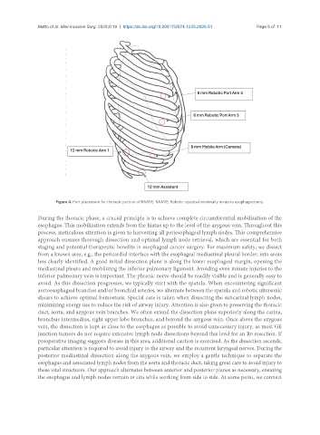
Figure 4. Port placement for thoracic portion of RAMIE. RAMIE: Robotic-assisted minimally invasive esophagectomy.
During the thoracic phase, a crucial principle is to achieve complete circumferential mobilization of the
esophagus. This mobilization extends from the hiatus up to the level of the azygous vein. Throughout this
process, meticulous attention is given to harvesting all periesophageal lymph nodes. This comprehensive
approach ensures thorough dissection and optimal lymph node retrieval, which are essential for both
staging and potential therapeutic benefits in esophageal cancer surgery. For maximum safety, we dissect
from a known area, e.g., the pericardial interface with the esophageal mediastinal pleural border, into areas
less clearly identified. A good initial dissection plane is along the lower esophageal margin, opening the
mediastinal pleura and mobilizing the inferior pulmonary ligament. Avoiding even minute injuries to the
inferior pulmonary vein is important. The phrenic nerve should be readily visible and is generally easy to
avoid. As this dissection progresses, we typically start with the spatula. When encountering significant
aortoesophageal branches and/or bronchial arteries, we alternate between the spatula and robotic ultrasonic
shears to achieve optimal hemostasis. Special care is taken when dissecting the subcarinal lymph nodes,
minimizing energy use to reduce the risk of airway injury. Attention is also given to preserving the thoracic
duct, aorta, and azygous vein branches. We often extend the dissection plane superiorly along the carina,
bronchus intermedius, right upper lobe bronchus, and beyond the azygous vein. Once above the azygous
vein, the dissection is kept as close to the esophagus as possible to avoid unnecessary injury, as most GE
junction tumors do not require extensive lymph node dissections beyond this level for an R0 resection. If
preoperative imaging suggests disease in this area, additional caution is exercised. As the dissection ascends,
particular attention is required to avoid injury to the airway and the recurrent laryngeal nerves. During the
posterior mediastinal dissection along the azygous vein, we employ a gentle technique to separate the
esophagus and associated lymph nodes from the aorta and thoracic duct, taking great care to avoid injury to
these vital structures. Our approach alternates between anterior and posterior planes as necessary, ensuring
the esophagus and lymph nodes remain in situ while working from side to side. At some point, we connect