
Page 13 - Read Online
P. 13
Page 4 of 11 Matto et al. Mini-invasive Surg. 2025;9:19 https://dx.doi.org/10.20517/2574-1225.2025.51
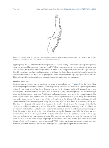
Figure 3. Completed true EEA (intrathoracic esophagogastric) with the restapled area forming a uniform 3-cm-wide conduit. Arrows
indicate the location of the 3-cm-wide conduit. EEA: End-to-end anastomosis.
a pyloroplasty. To conclude the abdominal portion, we place a feeding jejunostomy tube laparoscopically,
using our standard laparoscopic J-tube approach . While some surgeons are performing the jejunostomy
[7]
with the robot, we prefer a laparoscopic approach. Prior to the completion of the abdominal phase of the
RAMIE procedure, we place marking stitches to indicate the desired position of the lower border of the
newly created conduit relative to the diaphragmatic hiatus, in order to avoid pulling up too much conduit.
The marking stitch has been sufficient for accurate positioning in most circumstances.
Thoracic approach
For the thoracic portion, we use 4 robotic ports and 1 non-robotic port [Figure 4] for the chest. After
positioning the patient in the left lateral decubitus position, we access the thoracic cavity through the third
or fourth intercostal space. We chose this site to avoid the diaphragm, and it will ultimately serve as a
robotic port. Once the thoracic operative field is established, any adhesions present are divided using a
video-assisted thoracoscopic surgery (VATS) approach, enabling safe placement of the remaining ports. The
8 mm robotic camera port is placed over the ninth rib in the eighth intercostal space, along the mid-axillary
line, under direct vision. The next 8 mm robotic port is placed in the fifth or sixth intercostal space (at least
two interspaces above the camera port) along the same line, which is near the mid- to anterior axillary line.
The final robotic port, a 12 mm port, is placed in the ninth to tenth intercostal space, posterior to the
camera port and at least 8 cm away from it. The final location of all robotic ports may be adjusted based on
the patient’s body habitus. In addition, it is important to maintain close to 8 cm between each robotic port.
From the patient’s right to left, robotic arm 1 has the force bipolar (later exchanged for the robotic stapler),
robotic arm 2 holds the 30-degree robotic camera, arm 3 has either the ultrasonic shear or an energy
dissector, and arm 4 has an atraumatic grasper. The assistant port is placed between the inferior working
ports and as low as the costal margin-diaphragm interface will allow. This 12 mm port serves as a second
12 mm robotic port but is mostly used as a standard VATS port for suctioning, suture and sponge insertion,
and small specimen retrievals. We use it for stapling if the angle appears superior to robotic arm 1.