
Page 56 - Read Online
P. 56
Sakane Anatomical relationship between Kambin’s triangle and ENR
In 1995, Mirkovic et al. clarified that a working cannula Arslan et al. [13] also showed anatomical variation in
[8]
could be safety placed in line with the medial one- the distance between the ENR and pedicle and the
third of corresponding pedicle. Min et al. reported an height and width of intervertebral foramen from L1-L2
[9]
average distance of 11.6 mm between the ENR and to L5-S1 in 14 male formalin-fixed cadavers.
the superior articulating process. Hoshide et al. also
[10]
measured the height and width of 16 Kambin’s safety ENR INJURY
triangles from 2 cadavers by closely penetrating
intervertebral discs using a standard posterolateral ENR injury is the most devastating complication of
[3]
approach with a Kirschner wire under fluoroscopic transforaminal PELD. In 2002, Yeung and Tsou
assistance. At the time of open dissection, there was reported on surgical outcomes and complications.
no ENR injury from the wire insertion. They showed The rate of postoperative dysesthesia (POD) was
[4]
averaged Kambin’s safety zone areas of 60, 71.5, 1.9% (6/307) with a 6-mm scope. Ruetten et al.
93.5, and 108 mm at L1-L2, L2-L3, L3-L4, and L4- reported POD in 1 (1.8%) out of 41 patients with an
2
L5 levels, respectively. Hardenbrook et al. also 8-mm cannula under general anesthesia. Ahn et al. [14,15]
[11]
analyzed Kambin’s safety zone areas by removing reported that POD occurred as a complication of
the top of a superior facet from 8 fresh-frozen female PELD under local anesthesia and sedation in 4.7%
cadaveric specimens, and reported averaged areas of of recurrent herniated cases and in 6.7% of upper
115, 120, 119, and 116 mm at L1-L2, L2-L3, L3-L4, lumbar lesion cases. In their early case series of
2
transforaminal PELD with an 8-mm diameter scope,
and L4-L5 levels, respectively. They concluded that Abe et al. [16] reported that 2 (9.6%) and 4 (19%) of
Kambin’s working triangle was a relatively large area 22 patients experienced POD after surgery under
for minimally invasive transforaminal interbody fusion. general and local anesthesia, respectively. Although
On the other hand, Ozer et al. performed both they used a contrast material injection technique in
[12]
cadaveric measurements and surgical observations the epidural space to determine the ENR anatomy
of Kambin’s safety zone. They observed only 17.6% during surgery, it did not prevent nerve irritation. [17]
and 10.8% of “wide” safety zones of cadaveric
measurements and surgical observations, respectively Choi et al. evaluated clinical-radiological features
[18]
and concluded that there were large variations in indicating a risk of root injuries for proposed
Kambin’s triangle. Furthermore, there was no space transforaminal endoscopic discectomy. In their
inside the triangle in approximately one-third of L2-L5 retrospective analysis of 233 patients treated
in cadaveric (15/48) and surgical specimens (11/34). with PELD for lumbar disc herniation, 20 (4.7%)
They suggested using a partial superior facetectomy patients exhibited postoperative exiting root-related
to avoid ENR injury [Figure 2]. dysesthesias or motor weakness. They did not
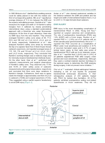
Figure 1: Kambin’s safety triangle (shaded area) at L4/L5
100 Mini-invasive Surgery ¦ Volume 1 ¦ September 30