
Page 29 - Read Online
P. 29
Ohara et al. Percutaneous endoscopic lumbar laminectomy
A B A B
C C
D D
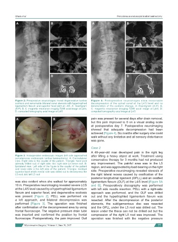
Figure 2: Preoperative neuroimages reveal degenerative lumbar Figure 4: Postoperative neuroimages show reasonable
scoliosis and remarkable bilateral canal stenosis with hypertrophied decompression of the spinal canal at the L4/5 level and no
ligamentum flavum and superior facet joint at L4/5. A: Roentgram deterioration of the scoliotic change. A: Roentgram (A-P); B,
(A-P); B, C: magnetic resonance imaging T2WI axial image at L4/5; C: magnetic resonance imaging T2WI axial image at L4/5; D:
D: computed tomography axial image at L4/5 computed tomography axial image at L4/5
A B pain was present for several days after drain removal,
but this pain improved to 0 on a visual analog scale
at postoperative day 7. Postoperative neuroimaging
showed that adequate decompression had been
Cranial
achieved [Figure 4]. Six months after surgery she could
walk without any limitation and all sensory disturbance
Cranial
was gone.
Caudal
Case 2
Left
A 48-year-old man developed pain in the right leg
Figure 3: Intraoperative endoscopic images (left side approached after lifting a heavy object at work. Treatment using
percutaneous endoscopic lumbar laminectomy). A: Contralateral conservative therapy for 3 months had not produced
view. Right side is the caudal of the patient. Triangle: facet joint
(already drilled out) of right side; dot: right side L5 nerve root. B: any improvement. The painful area was in the L5
Ipsilateral view. Left side of the figure is the caudal of the patient region, and was aggravated by load-bearing on the right
and lower side is the left side of the patient. Triangle: residual
superior facet which medial side was drilled out to decompress the side. Preoperative neuroimaging revealed stenosis of
L5 root; dot: left L5 root the right lateral recess caused by ossification of the
posterior longitudinal ligament (OPLL) and an ossified
was also evident when she walked for approximately ligamentum flavum (OLF) at the L4/5 level [Figure 5A
10 m. Preoperative neuroimaging revealed severe LCS and B]. Preoperatively discography was performed
at the L4/5 level caused by a hypertrophied ligamentum with left side needle insertion. PELL with a right-side
flavum and superior facet, and degenerative scoliosis approach was performed, and the OLF was drilled
was present [Figure 2]. PELL was performed via out and the hypertrophied ligamentum flavum was
a left approach, and bilateral decompression was resected. After the decompression of the posterior
performed [Figure 3]. The operation was finished elements, the subligamentous disc was resected
after confirmation of the decompressed area by using and the OPLL under the L5 root was drilled out. The
frontal fluoroscope. The negative pressure drain tube lesion under the theca can not be drilled out but the
was inserted and confirmed the position by frontal compression of the right L5 root was improved. The
fluoroscope. Postoperatively, the pain improved. Dull operation was finished with the negative pressure
Mini-invasive Surgery ¦ Volume 1 ¦ June 30, 2017 77