
Page 28 - Read Online
P. 28
Ohara et al. Percutaneous endoscopic lumbar laminectomy
A B
C D
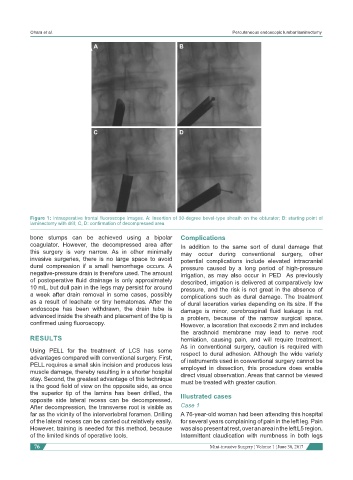
Figure 1: Intraoperative frontal fluoroscope images. A: Insertion of 30-degree bevel-type sheath on the obturator; B: starting point of
laminectomy with drill; C, D: confirmation of decompressed area
bone stumps can be achieved using a bipolar Complications
coagulator. However, the decompressed area after In addition to the same sort of dural damage that
this surgery is very narrow. As in other minimally may occur during conventional surgery, other
invasive surgeries, there is no large space to avoid potential complications include elevated intracranial
dural compression if a small hemorrhage occurs. A pressure caused by a long period of high-pressure
negative-pressure drain is therefore used. The amount irrigation, as may also occur in PED As previously
of postoperative fluid drainage is only approximately described, irrigation is delivered at comparatively low
10 mL, but dull pain in the legs may persist for around pressure, and the risk is not great in the absence of
a week after drain removal in some cases, possibly complications such as dural damage. The treatment
as a result of leachate or tiny hematomas. After the of dural laceration varies depending on its size. If the
endoscope has been withdrawn, the drain tube is damage is minor, cerebrospinal fluid leakage is not
advanced inside the sheath and placement of the tip is a problem, because of the narrow surgical space.
confirmed using fluoroscopy. However, a laceration that exceeds 2 mm and includes
the arachnoid membrane may lead to nerve root
RESULTS herniation, causing pain, and will require treatment.
As in conventional surgery, caution is required with
Using PELL for the treatment of LCS has some respect to dural adhesion. Although the wide variety
advantages compared with conventional surgery. First, of instruments used in conventional surgery cannot be
PELL requires a small skin incision and produces less employed in dissection, this procedure does enable
muscle damage, thereby resulting in a shorter hospital direct visual observation. Areas that cannot be viewed
stay. Second, the greatest advantage of this technique must be treated with greater caution.
is the good field of view on the opposite side, as once
the superior tip of the lamina has been drilled, the Illustrated cases
opposite side lateral recess can be decompressed.
After decompression, the transverse root is visible as Case 1
far as the vicinity of the intervertebral foramen. Drilling A 76-year-old woman had been attending this hospital
of the lateral recess can be carried out relatively easily. for several years complaining of pain in the left leg. Pain
However, training is needed for this method, because was also present at rest, over an area in the left L5 region.
of the limited kinds of operative tools. Intermittent claudication with numbness in both legs
76 Mini-invasive Surgery ¦ Volume 1 ¦ June 30, 2017