
Page 23 - Read Online
P. 23
Ohmori et al. FPCF for bony stenosis
DISCUSSION
Full-endoscopic spinal surgery, which is called
percutaneous endoscopic spinal surgery, was first
reported by Mayer and Brock [21] for the treatment of
lumbar disc herniation. Since then, surgeons have
developed a percutaneous endoscopic lumbar
discectomy through a transforaminal approach. [22-24]
In 2010, Choi et al. [25] devised a new technique that
approached the disc herniation through an interlaminar
window. Dezawa and Sairyo [26] further evolved the
procedure using a high-speed drill. Advances in the
interlaminar approach procedure have facilitated the
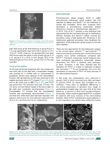
Figure 4: Postoperative computer tomography shows that vertical
fracture line is on the medial side on C6 vertebral lamina (arrow). R: application of full-endoscopic spinal surgery to cervical
right side spine disease.
pain VAS score at the final follow-up in group H (2.9 ± There are two approaches for full-endoscopic surgery
1.4) was significantly lower than that in group S (12 ± in the cervical spine: anterior [27,28] and posterior. [17-20]
16.4) (P < 0.05). However, the postoperative arm pain Anterior percutaneous endoscopic cervical discectomy
VAS scores were the same in both groups (group H 14 requires more careful techniques compared with
± 21, group S 14 ± 18.6). In both groups, 52% of the FPCF. [28] Therefore, endoscopic spinal surgeons who
facet joint (group H 52 ± 8.5%, group S 52 ± 6.7%) was have performed percutaneous endoscopic lumbar
resected. discectomy find FPCF a relatively easy technique
to learn. However, it has been reported that the
Case presentation indication for FPCF is limited to treatment of lateral
An 84-year-old female presented with very severe arm disc herniation. [17-20] To our knowledge, this is the first
and neck pain on the right side. Conservative therapy description of outcomes of FPCF for bony stenosis of
was pursued for 3 months with no improvement in the intervertebral foramen.
symptoms. Severe bony stenosis of the intervertebral
foramen with spondylosis at C5/6 was observed on the In this study, two complications were observed in
sagittal and axial views of CT images [Figure 5A and B] groupH, both of which occurred soon after we began
and on the axial view on MRI [Figure 5C], and FPCF to perform FPCF for lateral disc herniation at our
was performed. The operation time was 113 min. The institution. There were no significant differences in
C6 nerve root and lateral margin of the dura mater on the clinical parameters of operation time, length of
the right were completely decompressed [Figure 6]. postoperative hospital stay, arm pain VAS at the final
Postoperative CT showed that the intervertebral follow-up, or percent of facet joint resection between
foramen was successfully decompressed [Figure 7] the groups. These results suggest that FPCF is suitable
and that 42% of the facet joint had been resected. The for patients with bony stenosis of the intervertebral
patient’s VAS scores for neck and pain improved from foramen. When FPCF is performed in these patients, it
45.2 to 10.1 and from 63.4 to 5.2, respectively. is very important to be careful when drilling the lamina
A B C
Figure 5: (A) Sagittal computer tomography (CT) reveals that the intervertebral foramen is markedly narrowed on the right side at C5/6
(arrow); (b) foraminal stenosis and deformity of the facet joint on the right side are also observed in axial CT image (arrow); (C) axial magnetic
resonance imaging image also reveals that the foraminal stenosis on the right side is severe compared to that on the left (arrow). R: right side
66 Mini-invasive Surgery ¦ Volume 1 ¦ June 30, 2017