
Page 21 - Read Online
P. 21
Ohmori et al. FPCF for bony stenosis
INTRODUCTION
The upper extremity pain experienced by patients
with cervical radiculopathy is commonly caused by
either lateral cervical disc herniation or stenosis of the
intervertebral foramen due to a bone spur resulting
from spondylosis. Surgical treatment of cervical
radiculopathy can be divided into two procedures:
anterior cervical decompression and fusion [1-4] or
posterior foraminotomy. [5-9] The latter option involves
three types of procedures: open, [5-9] microscopic [10,11]
and micro-endoscopic surgery. [12-16]
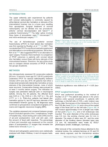
Figure 1: Percentage of resection of the facet joint was calculated
The use of full-endoscopic posterior cervical by Y/X × 100% which was measured on the coronal plane on
foraminotomy (FPCF) to treat lateral disc herniation postoperative computer tomography scans
was first reported by Ruetten et al. [17,18] in 2007. They
concluded that FPCF is a sufficient and safe supplement A B C
and alternative to conventional procedures. Since then,
Kim et al. [19,20] also suggested FPCF is an alternative to
open surgery. However, there has been no comparison
of FPCF outcomes in patients with cervical lateral
disc herniation versus those with bony stenosis of the
intervertebral foramen. Therefore, the aim of this study
was to compare clinical outcomes of FPCF in these
two groups of patients.
METHODS
We retrospectively assessed 59 consecutive patients Figure 2: Intraoperative images determining the location of the skin
[45 men, 14 women; mean age 53.7 (30-81) years] who incision. Location of the intervertebral disc at the C5/6 level (A) and
underwent FPCF for cervical radiculopathy between the medial edge of the facet joint (B) were marked as lines under
October 2014 and July 2016. All patients had either a intraoperative fluoroscopy. A small skin incision (C) approximately
8 mm in length is made at the intersection (arrow)
single-level symptomatic lateral disc herniation or bony
stenosis of the intervertebral foramen, none of which Statistical significance was defined as P < 0.05 (two-
were recurrent. Conservative therapy was pursued for sided).
at least 3 months before surgery. The indication for
surgery was persistent radicular pain or neurological FPCF surgical technique
deficits. Among the 59 patients, the affected level FPCF was performed according to the method of
was C4/5 (n = 16), C5/6 (n = 25), C6/7 (n = 16), and Ruetten et al. [17] The patient was placed in the prone
C7/T1 (n = 2). Thirty-four patients had lateral disc position under general anesthesia. In patients with
herniation (group H), and 25 had bony stenosis of the pathology on the left side of C5/6, a skin incision was
intervertebral foramen (group S). All diagnoses were made under fluoroscopy at the intersection of a line at
confirmed on preoperative computed tomography (CT) the C5/6 disc level [Figure 2A] and a line at the medial
scans and magnetic resonance imaging (MRI). edge of the facet joint on the left side [Figure 2B]. A
skin incision approximately 8 mm in length was made
The variables assessed and compared between groups at that point [Figure 2C], and a full endoscope was
H and S included operative time, complications, length inserted. The outer diameter of the entire endoscope
of hospital stay, visual analog pain scale (VAS) scores was 6.9 mm; the working channel was 4.2 mm in
of pre-and postoperative neck and arm pain, and diameter. The angle of vision was 25°, and the outer
the amount of facet joint resection. The percentage diameter of the working sleeve (beveled type) was
of facet joint resection was measured on the coronal 7.9 mm. All instruments were made by WOLF
plane of postoperative CT images that revealed the (RIWOspine GmbH, Knittlingen, Germany).
widest bone removal [Figure 1].
After removal of the connective tissue attached to the
Clinical and radiographic parameters were statistically vertebral lamina, the vertebral laminae at C5 and C6
analyzed with Mann-Whitney U and chi-square tests. were clearly exposed, and the interlaminar window
64 Mini-invasive Surgery ¦ Volume 1 ¦ June 30, 2017