
Page 59 - Read Online
P. 59
Page 4 of 10 Ditonno et al. Mini-invasive Surg 2023;7:36 https://dx.doi.org/10.20517/2574-1225.2023.62
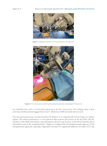
Figure 2. Docking of the Da Vinci SP® surgical system. SP: Single port.
Figure 3. The SP access port with the multichannel port and robotic instruments. SP: Single port.
the medullary layer and a 0 Vycril interrupted suture for the cortical layer. The bulldog clamp is then
removed, and the specimen bagged (Endo Catch™, Medtronic, Dublin, Ireland) and extracted.
The retroperitoneal access was first described by Maurice et al. using the SP1098 prototype on cadaver
models. The authors performed a 2.5 cm transverse skin, anterior and inferior to the tip of the 12th rib.
Division of the flank musculature and subsequent exposure and incision of the thoracolumbar fascia
[12]
allowed for access to the retroperitoneum . Bang et al. compared the retroperitoneoscopic approach to the
transperitoneal approach, reporting comparable outcomes. No significant difference in terms of OT and