
Page 58 - Read Online
P. 58
Ditonno et al. Mini-invasive Surg 2023;7:36 https://dx.doi.org/10.20517/2574-1225.2023.62 Page 3 of 10
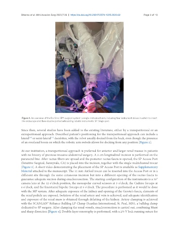
Figure 1. An overview of the Da Vinci SP® surgical system: a single instrument arm, including four instrument drives in which to insert
the endoscope and three double-jointed articulating robotic instruments. SP: Single port.
Since then, several studies have been added to the existing literature, either by a transperitoneal or an
extraperitoneal approach. Described patient’s positioning for the transperitoneal approach can include a
[10]
lateral or semi-lateral decubitus, with the robot usually docked from the back, even though the presence
[11]
of an overhead boom on which the robotic arm swivels allows for docking from any position [Figure 2].
At our institution, a transperitoneal approach is preferred for anterior and larger renal masses in patients
with no history of previous invasive abdominal surgery. A 3 cm longitudinal incision is performed on the
pararectal line. After rectus fibers are spread and the posterior rectus fascia is opened, the SP Access Port
(Intuitive Surgical, Sunnyvale, CA) is placed into the incision, together with the single multichannel trocar
[Figure 3]. A short video demonstrating the placement of the SP Access Port is available as Supplementary
Material attached to the manuscript. The 12 mm AirSeal trocar can be inserted into the Access Port or in a
different site through the same cutaneous incision but into a different opening of the rectus fascia to
guarantee adequate suction during enucleoresection. The starting configuration of the instruments is a 30°
camera lens at the 12 o’clock position, the monopolar curved scissors at 3 o’clock, the Cadiére forceps at
6 o’clock, and the fenestrated bipolar forceps at 9 o’clock. The procedure is performed as it would be done
with the MP system. After adequate exposure of the kidney and opening of the Gerota’s fascia, elements of
the renal pedicle are exposed. Isolation of the renal artery and vein is achieved, and adequate identification
and exposure of the renal mass is obtained through defatting of the kidney. Artery clamping is achieved
with the SCANLAN® Reliance Bulldog LP Clamp (Scanlan International, St. Paul, MN), a bulldog clamp
dedicated to SP surgery. After clamping the renal vessels, enucleoresection is carried out, combining blunt
and sharp dissection [Figure 4]. Double-layer renorraphy is performed, with a 2/0 V lock running suture for