
Page 39 - Read Online
P. 39
Page 6 of 9 Ciria et al. Mini-invasive Surg 2024;8:10 https://dx.doi.org/10.20517/2574-1225.2023.126
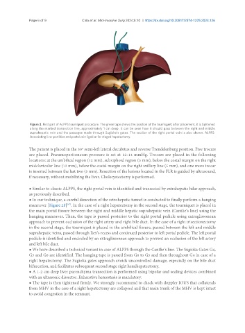
Figure 3. First part of ALPPS tourniquet procedure. The green tape shows the position of the tourniquet; after placement, it is tightened
along the marked transection line, approximately 1 cm deep. It can be seen how it should pass between the right and middle
suprahepatic vein and the passages made through Sugioka’s gates. The section of the right portal vein is also shown. ALPPS:
Associating liver partition and portal vein ligation for staged hepatectomy.
The patient is placed in the 30º semi-left lateral decubitus and reverse Trendelenburg position. Five trocars
are placed. Pneumoperitoneum pressure is set at 12-14 mmHg. Trocars are placed in the following
locations: at the umbilical region (12 mm), subxiphoid region (5 mm), below the costal margin on the right
midclavicular line (12 mm), below the costal margin on the right axillary line (5 mm), and one more trocar
is inserted between the last two (5 mm). Resection of the lesions located in the FLR is guided by ultrasound,
if necessary, without mobilizing the liver. Cholecystectomy is performed.
● Similar to classic ALPPS, the right portal vein is identified and transected by extrahepatic hilar approach,
as previously described.
● In our technique, a careful dissection of the retrohepatic tunnel is conducted to finally perform a hanging
maneuver [Figure 2F] . In the case of a right hepatectomy in the second stage, the tourniquet is placed in
[47]
the main portal fissure between the right and middle hepatic suprahepatic vein (Cantlie’s line) using the
hanging maneuver. Then, the tape is passed posterior to the right portal pedicle using extraglissonean
approach to prevent occlusion of the right artery and right bile duct. In the case of a right trisectionectomy
in the second stage, the tourniquet is placed in the umbilical fissure, passed between the left and middle
suprahepatic veins, passed through Rex’s recess and continued posterior to left portal pedicle. The left portal
pedicle is identified and encircled by an extraglissonean approach to prevent an occlusion of the left artery
and left bile duct.
● We have described a technical variant in case of ALPPS through the Cantlie’s line. The Sugioka Gates G4,
G5 and G6 are identified. The hanging tape is passed from G6 to G5 and then throughout G4 in case of a
right hepatectomy. The Sugioka gates approach avoids uncontrolled damage, especially on the bile duct
bifurcation, and facilitates subsequent second stage right hemihepatectomy.
● A 1-2 cm deep liver parenchyma transection is performed using bipolar and sealing devices combined
with an ultrasonic dissector. Exhaustive hemostasis is mandatory.
● The tape is then tightened firmly. We strongly recommend to check with doppler IOUS that collaterals
from MHV in the case of a right hepatectomy are collapsed and that main trunk of the MHV is kept intact
to avoid congestion in the remnant.