
Page 37 - Read Online
P. 37
Page 4 of 9 Ciria et al. Mini-invasive Surg 2024;8:10 https://dx.doi.org/10.20517/2574-1225.2023.126
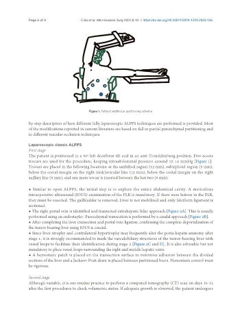
Figure 1. Patient and trocar positioning scheme.
by-step description of how different fully laparoscopic ALPPS techniques are performed is provided. Most
of the modifications reported in current literature are based on full or partial parenchymal partitioning and
in different vascular occlusion techniques.
Laparoscopic classic ALPPS
First stage
The patient is positioned in a 30º left decubitus tilt and in an anti-Trendelenburg position. Five access
trocars are used for the procedure, keeping intraabdominal pressure around 12-14 mmHg [Figure 1].
Trocars are placed in the following locations: at the umbilical region (12 mm), subxiphoid region (5 mm),
below the costal margin on the right midclavicular line (12 mm), below the costal margin on the right
axillary line (5 mm), and one more trocar is inserted between the last two (5 mm).
● Similar to open ALPPS, the initial step is to explore the entire abdominal cavity. A meticulous
intraoperative ultrasound (IOUS) examination of the FLR is mandatory. If there were lesions in the FLR,
they must be resected. The gallbladder is removed. Liver is not mobilized and only falciform ligament is
sectioned.
● The right portal vein is identified and transected extrahepatic hilar approach [Figure 2A]. This is usually
performed using an endostapler. Parenchymal transection is performed by a caudal approach [Figure 2B].
● After completing the liver transection and portal vein ligation, confirming the complete deportalization of
the tumor-bearing liver using IOUS is crucial.
● Since liver atrophy and contralateral hypertrophy may frequently alter the porta hepatis anatomy after
stage 1, it is strongly recommended to mark the vasculobiliary structures of the tumor-bearing liver with
vessel loops to facilitate their identification during stage 2 [Figure 2C and D]. It is also advisable but not
mandatory to place vessel loops surrounding the right and middle hepatic veins.
● A hemostatic patch is placed on the transection surface to minimize adhesion between the divided
sections of the liver and a Jackson-Pratt drain is placed between partitioned livers. Hemostasis control must
be rigorous.
Second stage
Although variable, it is our routine practice to perform a computed tomography (CT) scan on days 10-14
after the first procedures to check volumetric status. If adequate growth is observed, the patient undergoes