
Page 72 - Read Online
P. 72
Ghaseminejad et al. J Transl Genet Genom 2022;6:111-25 https://dx.doi.org/10.20517/jtgg.2021.49 Page 117
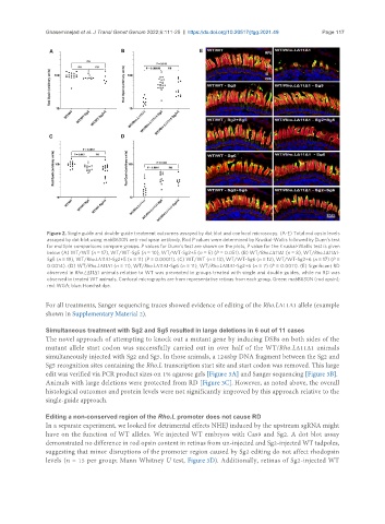
Figure 2. Single guide and double-guide treatment outcomes assayed by dot blot and confocal microscopy. (A-E) Total rod opsin levels
assayed by dot blot using mabB630N anti-rod opsin antibody. Rod P values were determined by Kruskal-Wallis followed by Dunn’s test
for multiple comparisons compare groups. P values for Dunn’s test are shown on the plots, P value for the Kruskal-Wallis test is given
below (A) WT/WT (n = 17), WT/WT-Sg5 (n = 10), WT/WT-Sg2+5 (n = 5) (P = 0.051). (B) WT/Rho.LΔ11Δ1 (n = 8), WT/Rho.LΔ11Δ1-
Sg5 (n = 19), WT/Rho.LΔ11Δ1-Sg2+5 (n = 11) (P = 0.00011). (C) WT/WT (n = 13), WT/WT-Sg6 (n = 12), WT/WT-Sg2+6 (n = 17) (P =
0.0014). (D) WT/Rho.LΔ11Δ1 (n = 11), WT/Rho.LΔ11Δ1-Sg6 (n = 11), WT/Rho.LΔ11Δ1-Sg2+6 (n = 7) (P = 0.0011). (E) Significant RD
observed in Rho.LΔ11Δ1 animals relative to WT was prevented in groups treated with single and double guides, while no RD was
observed in treated WT animals. Confocal micrographs are from representative retinas from each group. Green: mabB630N (rod opsin);
red: WGA; blue: Hoechst dye.
For all treatments, Sanger sequencing traces showed evidence of editing of the Rho.LΔ11Δ1 allele (example
shown in Supplementary Material 2).
Simultaneous treatment with Sg2 and Sg5 resulted in large deletions in 6 out of 11 cases
The novel approach of attempting to knock out a mutant gene by inducing DSBs on both sides of the
mutant allele start codon was successfully carried out in over half of the WT/Rho.LΔ11Δ1 animals
simultaneously injected with Sg2 and Sg5. In those animals, a 1248bp DNA fragment between the Sg2 and
Sg5 recognition sites containing the Rho.L transcription start site and start codon was removed. This large
edit was verified via PCR product sizes on 1% agarose gels [Figure 3A] and Sanger sequencing [Figure 3B].
Animals with large deletions were protected from RD [Figure 3C]. However, as noted above, the overall
histological outcomes and protein levels were not significantly improved by this approach relative to the
single-guide approach.
Editing a non-conserved region of the Rho.L promoter does not cause RD
In a separate experiment, we looked for detrimental effects NHEJ induced by the upstream sgRNA might
have on the function of WT alleles. We injected WT embryos with Cas9 and Sg2. A dot blot assay
demonstrated no difference in rod opsin content in retinas from un-injected and Sg2-injected WT tadpoles,
suggesting that minor disruptions of the promoter region caused by Sg2 editing do not affect rhodopsin
levels (n = 15 per group; Mann Whitney U test, Figure 3D). Additionally, retinas of Sg2-injected WT