
Page 48 - Read Online
P. 48
Page 55 Ryan et al. J Transl Genet Genom. 2025;9:48-61 https://dx.doi.org/10.20517/jtgg.2024.87
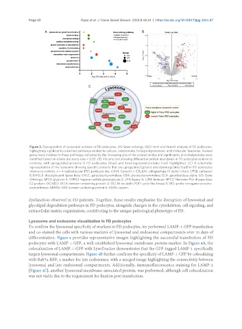
Figure 3. Dysregulation of lysosomal proteins in FD podocytes. (A) Gene ontology (GO) term enrichment analysis of FD podocytes,
highlighting significantly enriched pathways related to cellular components, biological processes, and molecular functions. Several
genes were involved in these pathways, indicated by the increasing size of the colored circles and significantly enriched proteins were
identified based on a false discovery rate < 0.05. (B) Volcano plot showing differential protein abundance in FD podocytes relative to
controls, with upregulated proteins in FD podocytes (blue) and downregulated proteins (red) highlighted. (C) A schematic
representation of the lysosome showing specific proteins that are upregulated (green) and downregulated (red) in FD podocytes
relative to controls. n = 4 replicates per iPSC-podocyte line. CAV1: Caveolin 1; COL6A1: collagen type IV alpha 1 chain; CTSB: cathepsin
B; DAGLB: diacylglycerol lipase beta; GALC: galactosylceramidase; GBA: glucosylceramindase; GLA: galactosidase alpha; GO: Gene
Ontology; GPC4: glypican 4; HSPG2: heparan sulfate proteoglycan 2; LIPA: lipase A; LUM: lumican; NPC2: Niemann-Pick disease type
C2 protein; OCIAD2: OCIA domain-containing protein 2; OCLN: occludin; PLK1: polo-like kinase 1; SRC: proto-oncogene tyrosine-
protein kinase; UBXN6: UBX domain-containing protein 6; VASN: vasorin.
dysfunction observed in FD patients. Together, these results emphasize the disruption of lysosomal and
glycolipid degradation pathways in FD podocytes, alongside changes in the cytoskeleton, cell signaling, and
extracellular matrix organization, contributing to the unique pathological phenotype of FD.
Lysosome and endosome visualization in FD podocytes
To confirm the lysosomal specificity of markers in FD podocytes, we performed LAMP-1-GFP transfection
and co-stained the cells with various markers of lysosomal and endosomal compartments over 20 days of
differentiation. Figure 4 provides representative images highlighting the successful transfection of FD
podocytes with LAMP-1-GFP, a well-established lysosomal membrane protein marker. In Figure 4A, the
colocalization of LAMP-1-GFP with LysoTracker demonstrates that the GFP-tagged LAMP-1 specifically
targets lysosomal compartments. Figure 4B further confirms the specificity of LAMP-1-GFP by colocalizing
with Rab7a-RFP, a marker for late endosomes, with a merged image highlighting the connectivity between
lysosomal and late endosomal compartments. Additionally, immunofluorescence staining for LAMP-2
[Figure 4C], another lysosomal membrane-associated protein, was performed, although cell colocalization
was not viable due to the requirement for fixation post transfection.