
Page 502 - Read Online
P. 502
Page 2 of 9 Wu et al. J Cancer Metastasis Treat 2020;6:40 I http://dx.doi.org/10.20517/2394-4722.2020.77
[1]
most common toxicities . Most immunotherapy-induced cutaneous toxicities are mild, such as non-
specific rashes or pruritus. However, some manifestations of skin irAEs can progress to high-grade and
[2]
potentially life-threatening situations, such as bullous pemphigoid (BP) . Since 2015, more than 40 cases
of immunotherapy-induced BP have been reported with the majority involving either the skin or mucous
membrane only. Presentations of both cutaneous and oral mucous BP are rare (less than seven cases).
Here, we report a severe, extensive (involving both skin and oral mucosa), refractory case of BP that
occurred 9 months after initiation of nivolumab in a patient with metastatic renal clear cell cancer. We
also summarise a list of selected case reports of checkpoint inhibitor-induced BP by literature review. We
highlight various presentations, investigations and management approaches of immunotherapy-induced
BP. Meantime, we would like to discuss the correlation of skin irAE incidence rate with immunotherapy
drug benefit and resistance.
CASE REPORT
A 66-year-old gentleman with no known history of autoimmune disease was diagnosed with right clear
renal cancer in 2014. He was initially treated with radical nephrectomy with excision of tumour thrombus
in the renal vein and retro-hepatic inferior vena cava. In March 2017, his cancer relapsed with a single
spinal metastasis, which was treated with excision of the intradural tumour and laminectomy and
radiotherapy as well, which resulted in complete neurological recovery. Post-radiotherapy, he opted for
active tyrosine kinase inhibitor, sunitinib, in light of relapsed renal cancer in his spine. In October 2017,
sunitinib was discontinued due to spontaneous haematoma in his right calf and thigh, which was likely due
to combination of sunitinib and low-molecular-weight heparin (LMWH) for his atrial fibrillation. He then
had a long period of surveillance, during which he gradually developed asymptomatic relapsed metastatic
renal cancer in his left adrenal gland, followed by relapsed cancer in his left kidney. In March 2019, he
commenced immunotherapy infusion with 2 weekly nivolumab, considering further disease progression
with multiple lung metastatic disease. His other medical history was only atrial fibrillation, for which he
was on LMWH and bisoprolol.
From August 2019, five months post-initiation of nivolumab, he developed a mild pruritic erythematous
skin rash scattered mainly over his chest and upper limbs. At that time, there were no blisters reported. He
continued nivolumab with the rash controlled mainly by topical steroids or short courses of oral steroids.
In November 2019, he developed large, tense, haemorrhagic blisters, which were exacerbated following a
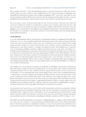
3-week holiday in Australia, leading to an emergency hospital admission. On clinical examination, he had
a severe widespread bullous eruption affecting his chest, abdomen, inner thighs and upper arms. Active
blisters and erosions were seen on the roof of his mouth and scrotal skin as well. Some blisters had burst
leaving severely eroded skin with signs of superimposed infection. Bullae at various stages were seen,
including intact ones, burst ones with an eroded base, and a few areas of early re-epithelialisation as Figure 1.
He had no involvement of the scalp region and external eye examination was normal.
An incisional skin biopsy from an active blister with haematoxylin and eosin examination revealed
epidermal hyperplasia and subepidermal splitting. There were few eosinophils, scattered plasma cells and
neutrophils with a subjacent chronic inflammation. Direct immunofluorescence (DIF) showed clearly
positive staining for IgG in the basement membrane zone, and a faint positive IgA staining. Subsequent
serum antibody analysis confirmed positive pemphigoid antibodies consistent with BP see Figures 2 and 3.
The patient was started immediately on intravenous methylprednisolone 1 mg/kg as well as flucloxacillin
for 4 days and then switched to oral prednisolone and flucloxacillin when no new blisters developed.
Most of the blisters gradually re-epithelialised within 4-6 weeks of oral steroids. Two months later, the
corticosteroid treatment was tapered. However, the blisters reappeared when prednisolone was stepped