
Page 658 - Read Online
P. 658
Page 10 of 14 Tafur et al. J Cancer Metastasis Treat 2018;5:xx I http://dx.doi.org/10.20517/2394-4722.2018.102
A MCT1 PR
siNT + + _ _ siNT + + _ _
siGPR81 _ _ + + siGPR81 _ _ + +
Tamoxifen _ + _ + Tamoxifen _ + _ +
B Ki67 CyclinB1
siNT + + _ _ siNT + + _ _
siGPR81 _ _ + + siGPR81 _ _ + +
Tamoxifen _ + _ + Tamoxifen _ + _ +
C D Cleaved PARP
siNT + + _ _ siNT + + _ _
siGPR81 _ _ + + siGPR81 _ _ + +
Tamoxifen _ + _ + Tamoxifen _ + _ +
cleaved PARP
β-actin
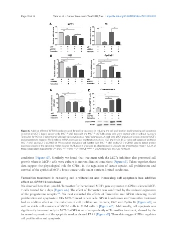
Figure 6. Additive effect of GPR81 knockdown and Tamoxifen treatment in reducing the cell proliferation and increasing cell apoptosis
in epithelial MCF-7 breast cancer cells. MCF-7-siNT (control) and MCF-7-siGPR81 cancer cells were treated with or without 1 μmol/L
Tamoxifen for 96 h in 3-dimensional Matrigel with physiological modified medium. A: real-time qPCR analysis of lactate importer MCT1;
and progesterone receptor PR; B: relative mRNA expression of proliferation markers: Ki67 and Cyclin B1; C: viable cell counts of epithelial
MCF-7-siNT and MCF-7-siGPR81; D: Western blot analysis of cell lysates from MCF-7-siNT and MCF-7-siGPR81 used to detect protein
expression levels of the apoptotic maker cleaved-PARP. β-actin was used as a loading control. Results are presented as mean ± S.E.M. of
three independent experiments *P < 0.05; **P < 0.01; ***P < 0.001; ****P < 0.0001 (ordinary one-way ANOVA)
conditions [Figure 5D]. Similarly, we found that treatment with the MCT1 inhibitor also prevented cell
growth when in MCF-7 cells were culture in nutrient-limited conditions [Figure 5E]. Taken together, these
data support the physiological role for GPR81 in the regulation of lactate uptake, cell proliferation and
survival of the epithelial MCF-7 breast cancer cells under nutrient-limited conditions.
Tamoxifen treatment in reducing cell proliferation and increasing cell apoptosis has additive
effect on GPR81 knockdown
We observed here that 1 µmol/L Tamoxifen further reduced MCT1 gene expression in GPR81-silenced MCF-
7 cells treated for 4 days [Figure 6A]. The effect of Tamoxifen was confirmed by the reduced expression
of the progesterone receptor . We next evaluated the effects of Tamoxifen and GPR81 silencing in cell
[35]
proliferation and apoptosis in ER+ MCF-7 breast cancer cells. GPR81 knockdown and Tamoxifen treatment
had an additive effect on the reduction of cell proliferation markers; Ki67 and Cyclin B1 [Figure 6B], as
well as viable cell numbers of MCF-7 cells in MPM culture [Figure 6C]. Additionally, cell apoptosis was
significantly increased only in MCF-7-siGPR81 cells independently of Tamoxifen treatment, showed by the
increased expression of the apoptotic marker cleaved PARP [Figure 6E]. These data suggest GPR81 regulates
cell proliferation and apoptosis.