
Page 654 - Read Online
P. 654
Page 6 of 14 Tafur et al. J Cancer Metastasis Treat 2018;5:xx I http://dx.doi.org/10.20517/2394-4722.2018.102
A E M B
GPR81
MCF-7
BT-474
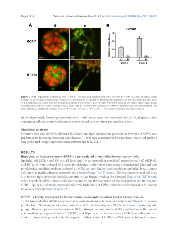
Figure 1. GPR81 expression of epithelial MCF-7 and BT-474 cells and mesenchymal MCF-7M and BT-474M in 3-dimensional Matrigel
culture. A: fluorescence microscopy images of 3-dimensional structures of proliferating epithelial (E) and mesenchymal (M) cells
in 3-dimensional Matrigel with physiological modified medium for 7 days. Green, Phalloidin staining of F-actin; red/orange, nuclei
counterstained with SYTOXTM orange nucleic acid (red); B: real-time qPCR analysis of GPR81 in epithelial (E) and mesenchymal (M)
cells. Results are presented as mean ± S.E.M. *P < 0.05; **P < 0.01; ***P < 0.001; ****P < 0.0001 (ordinary one-way ANOVA)
in the signal used. Resulting concentrations in millimolar were then exported into an Excel spread sheet
containing cellular counts to determine a normalized concentration per number of cells.
Statistical analysis
Ordinary one-way ANOVA followed by Sidak’s multiple comparison post-test or two-way ANOVA was
performed to determine statistical significance. P < 0.05 was considered to be significant. Statistical analysis
was performed using GraphPad Prism software (La Jolla, CA).
RESULTS
Endogenous lactate receptor GPR81 is upregulated in epithelial breast cancer cells
Epithelial (E) MCF-7 and BT-474 cell lines and the corresponding post-EMT mesenchymal (M) MCF-7M
and BT-474M were cultured in a more physiologically relevant system using 3-dimensional Matrigel and
physiological modified medium (referred as MPM culture). Under these conditions epithelial breast cancer
cells grew as tightly adherent spheroids for 1 week [Figure 1A; “E” boxes]. The two mesenchymal cell lines
also formed tight spheroids initially, but after 2 days began invading the Matrigel [Figure 1A; “M” boxes].
After 1 week of MPM culture, cells were examined for the expression of the endogenous lactate receptor
GPR81. Epithelial spheroids expressed relatively high levels of GPR81, whereas mesenchymal cells showed
no or very low expression [Figure 1B].
GPR81 is highly expressed in human hormone-receptor-positive breast cancer tissues
To determine whether GPR81 was present in human breast cancer tumors, we analyzed mRNA gene expression
profiles from 74 human breast cancer samples and 12 non-tumorigenic (NT) breast tissues [Figure 2A]. We
grouped these samples in non-tumorigenic (NT), estrogen receptor positive (ER ), amplification of the human
+
epidermal receptor growth factor 2 (HER2 ) and triple negative breast cancer (TNBC) according to their
+
clinical information provided by the supplier. Higher levels of GPR81 mRNA were noted in hormone-