
Page 553 - Read Online
P. 553
Morgan et al. J Cancer Metastasis Treat 2018;4:46 I http://dx.doi.org/10.20517/2394-4722.2018.19 Page 9 of 13
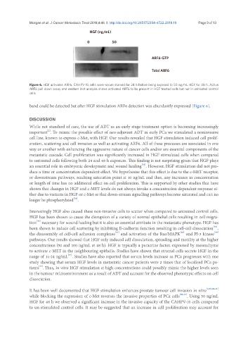
HGF (ng/mL)
0 50
ARF6-GTP
Total ARF6
Figure 6. HGF activates ARF6. CAHPV-10 cells were serum starved for 24 h before being exposed to 50 ng/mL HGF for 48 h. Active
ARF6 pull down assay and western blot analysis shows activated ARF6 to be present in HGF treated cells but not in untreated control
cells
band could be detected but after HGF stimulation ARF6 detection was abundantly expressed [Figure 6].
DISCUSSION
While not standard of care, the use of ADT as an early stage treatment option is becoming increasingly
[2]
important . To mimic the possible effect of neo-adjuvant ADT in early PCa we stimulated a noninvasive
cell line, known to express c-Met, with HGF. Our results revealed that HGF stimulation induced cell prolif-
eration, scattering and cell invasion as well as activating ARF6. All of these processes are associated in one
way or another with enhancing the aggressive nature of cancer cells and/or are essential components of the
metastatic cascade. Cell proliferation was significantly increased in HGF stimulated cells when compared
to untreated cells following both 24 and 48 h exposure. This finding is not surprising given that HGF plays
an essential role in embryonic development and wound healing . However, HGF stimulation did not pro-
[33]
duce a time or concentration dependent effect. We hypothesise that this effect is due to the c-MET receptor,
or downstream pathways, reaching saturation point at 10 ng/mL and thus, any increases in concentration
or length of time has no additional effect on cell proliferation. This is supported by other studies that have
shown that changes in HGF and c-MET levels do not always invoke a concentration-dependent response ei-
ther due to variants in HGF or c-Met or that down-stream signalling pathways become saturated and can no
longer be phosphorylated .
[34]
Interestingly HGF also caused these non-invasive cells to scatter when compared to untreated control cells.
HGF has been shown to cause the disruption of a variety of normal epithelial cells resulting in cell migra-
[35]
tion necessary for wound healing but it is also an essential attribute in the metastatic phenotype. HGF has
[36]
been shown to induce cell scattering by inhibiting E-cadherin function resulting in cell-cell dissociation ,
the disassembly of cell-cell adhesion complexes and activation of the Ras/MAPK and PI-3 kinase
[39]
[37]
[38]
pathways. Our results showed that HGF only induced cell dissociation, spreading and motility at the higher
concentrations (50 and 100 ng/mL at 48 h). HGF is typically a paracrine factor, expressed by mesenchyme
to activate c-MET in the neighbouring epithelia. Studies have shown that stromal cells secrete HGF in the
range of 14-24 ng/mL . Studies have also reported that serum levels increase as PCa progresses with one
[34]
study showing that serum HGF levels in metastatic cancer patients were 2 times that of localised PCa pa-
tients . Thus, in vitro HGF stimulation at high concentrations could possibly mimic the higher levels seen
[17]
in the tumour microenvironment as a result of ADT and account for the observed phenotypic effects on cell
dissociation.
It has been well documented that HGF stimulation enhances prostate tumour cell invasion in vitro [9,19,40,41]
while blocking the expression of c-Met reverses the invasive properties of PCa cells [40,42] . Using 50 ng/mL
HGF for 48 h we observed a significant increase in the invasive capacity of the CAHPV-10 cells compared
to un-stimulated control cells. It may be suggested that an increase in cell proliferation may account for