
Page 552 - Read Online
P. 552
Page 8 of 13 Morgan et al. J Cancer Metastasis Treat 2018;4:46 I http://dx.doi.org/10.20517/2394-4722.2018.19
A
B
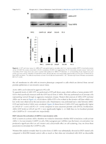
Figure 5. A: GST pull down assay for ARF6-GTP was performed on prostate cells from normal prostate (PNT2), localised prostate
cancer (CAHVP-10), weakly metastatic (LNCaP), moderately (DU145) and aggressively (PC-3) metastatic prostate cancer. Twenty five
microlitres of GST assay elute was used in western blot analysis for ARF6-GTP detection. A: corresponding 25 µL aliquot from total
protein lysis was used for detection of total ARF6 protein; B: band density was measured by densitometry and the ratios of ARF6-GTP to
total ARF6 are shown. The data are expressed as mean (from triplicate experiments) ± SD. *Denotes significant difference compared to
PNT2 (P < 0.05)
the cell membrane in cells with an invasive phenotype compared to cells which are derived from normal
prostate epithelium or non-invasive cancer tissue.
Active ARF6 can be detected in aggressive PCa cells
To quantify levels of ARF-GTP, we performed a GTP pull down assay, which utilises a fusion protein GST-
GGA3 that specifically interacts with the GTP bound form of ARF6. This was performed on all prostate cells
representing normal through to aggressive metastatic disease. Western blot analysis of ARF6-GTP and total
ARF6 can be seen in Figure 5A, which shows ARF6-GTP to be evident in the invasive cell lines whilst very
low levels were observed in the non-invasive cells. Densitometry was performed and a ratio between ARF6-
GTP and total levels of ARF6 were calculated. Figure 5B shows levels of ARF6-GTP were significantly higher
in LNCaP (P < 0.033) and PC3 (P < 0.034) compared to non-cancerous prostate cells (PNT2). Furthermore,
ARF6-GTP levels in LNCaP and PC-3 were significantly higher (> 2.5 fold) than in the localised prostate
cells CAHPV10 (P < 0.044 and 0.001 respectively).
HGF induces the activation of ARF6 in non-invasive cells
HGF is known to activate ARF6, therefore we wished to determine whether HGF stimulation could activate
ARF6 in the non-invasive CAHPV-10 cells. Fifty nanograms per millilitre was the lowest concentration that
produced a significant effect on cell invasion and a discernable effect on cell scattering, thus, we chose these
parameters to stimulate the CAHPV10 cells.
Western blot analysis revealed that the active form of ARF6 was substantially elevated in HGF treated cells
compared to BSA/PBS treated control cells so much so that when not stimulated with HGF no discernible