
Page 549 - Read Online
P. 549
Morgan et al. J Cancer Metastasis Treat 2018;4:46 I http://dx.doi.org/10.20517/2394-4722.2018.19 Page 5 of 13
A B
Absorbance (492 nm) Absorbance (492 nm)
HGF (ng/mL)
HGF (ng/mL)
C
Relative fluorescence (485/520 nmol/L)
HGF (ng/mL)
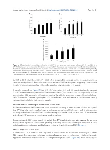
Figure 1. HGF significantly increased the proliferation of CAHPV-10 non-invasive prostate cancer cells over (A) 24 h and (B) 48 h.
CAHPV-10 cells were serum starved for 24 h before exposure to HGF. The data are expressed as mean ± standard deviation (SD) (from
4 replicates repeated in triplicate). *Denotes significant difference between treated and control cells (P < 0.05); C: HGF significantly
increased the invasive capacity of CAHPV-10 cells. Cells were seeded onto an artificial basement membrane extract in transwell inserts
and serum starved for 24 h before treatment with HGF for 48 h. The data are expressed as mean (from triplicate experiments) ± SD.
*Denotes significant difference between treated and control cells (P < 0.05). HGF: hepatocyte growth factor
by HGF at 24 (P < 0.001) and 48 h (P < 0.001) when compared to untreated control cells, yet interestingly
there was no significant difference between concentrations of HGF or time points, suggesting that the c-Met
receptor or downstream signaling pathways have reached saturation point at 10 ng/mL.
It can also be seen from Figure 1C that 48 h HGF stimulation at 50 and 100 ng/mL significantly increased
CAHPV-10 invasion through an artificial basement membrane (P < 0.042 and P < 0.019 respectively) with an
approximate 2-fold increase in cell numbers crossing the artificial membrane compared to untreated con-
trols. These results show that exposing PCa cells, derived from a localised tumour, to HGF increases not only
their proliferation but also their invasive capacity.
HGF induced cell scattering in non-invasive cancer cells
To determine whether HGF stimulation could induce cell scattering in a non-invasive cell line, we exposed
CAHPV-10 cells (grown in small colonies) to various concentrations of HGF over 24 and 48 h time period.
As many studies have shown HGF induce cell scattering in DU145 PCa cells, we also used DU145 cells with
and without HGF exposure as a positive and negative controls.
Concentrations of HGF ranged from 0-100 ng/mL. CAHPV-10 cells treated over a 24 h period did not show
any significant signs of cell dissociation, spreading or motility. In contrast, following 48 h exposure to HGF,
cell dissociation, spreading and motility became evident at 50 and 100 ng/mL HGF [Figure 2].
ARF6 is expressed in PCa cells
A role for the GTPase ARF6 has been implicated in several cancers but information pertaining to its role in
PCa is scant. Gene expression analysis on prostate cells derived from normal prostate epithelium through to
aggressive, metastatic disease revealed ARF6 to be present in prostate cells [Figure 3A]. There was no signifi-