
Page 551 - Read Online
P. 551
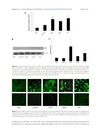
Morgan et al. J Cancer Metastasis Treat 2018;4:46  I  http://dx.doi.org/10.20517/2394-4722.2018.19                        Page 7 of 13

                              A


               B                                          C


               Figure 3. A: Gene expression analysis shows ARF6 is expressed in prostate cells derived from normal tissue (PNT2), localised prostate
               cancer (CAHVP-10), weakly metastatic (LNCaP), moderately (DU145) and aggressively (PC-3) metastatic prostate cancer tissue. ARF6
               expression was similar between PNT2 and CAHPV-10. A significant increase in expression was observed in LNCaP, DU145 and PC-3
               compared to CAHPV-10. The data are expressed as mean (from triplicate experiments), relative to PNT2, ± SD. *Denotes significant
               difference compared to PNT2 (P < 0.05); B: western blot analysis for total ARF6 protein; C: densitometry data is presented relative to the
               level of β-actin. The data are expressed as mean (from triplicate experiments) ± SD


                     PNT2                                             CAHPV-10                                      LNCaP                                     DU145                                            PC3
               Figure 4. Inactive ARF6 localises to the cytosol and endosomes and when activated translocates to the plasma membrane. Representative
               immunofluorescence images show cells stained with rabbit anti-ARF6 exhibit a stronger signal intensity at the plasma membrane in the
               metastatic cell lines (LNCaP, DU145, PC3) than those derived from normal tissue (PNT2) and localised prostate cancer (CAHPV-10).
               Corresponding negative controls are shown in the bottom row

               periphery in all cell types. In PNT2 cells, ARF6 staining intensity was very weak but intensity increased with
               progression to an aggressive phenotype suggesting higher levels of the activated form of ARF6 is present at