
Page 115 - Read Online
P. 115
Youbi et al. Management of choroidal metastasis using external beam radiotherapy
(n = 22) or bilateral (n = 6). The inaugural symptoms raison why patients should systematically benefit from
were variable: decreased visual acuity, visual field a complete specialized ophthalmologic examination,
amputation with scotoma, photophobia, myodesopsia prior to the initiation of a treatment. According to some
[Table 2]. authors, bilateral involvement is associated with a
shorter likelihood of survival. On ophtalmoscopy,
[9]
At the end of the irradiation, 13 patients (46%) showed choroidal metastases appear as flat orange lesions
an improvement in ophthalmologic symptoms. For located most often at the posterior pole of the eye,
the others, a stabilization of the symptoms was noted which can induce focal retinal detachment. Anterior
(n = 15). No patients showed visual degradation. No or posterior uveitis may sometimes be associated.
acute or late grade 2-3 toxicity was objectived. The Mode A (Amplitude) and B (Brightness) ultrasound
histological type was not significantly correlated with as well as fluorescein angiography can assist in
the response (P = 0.5) according to Fisher’s exact test. diagnosis. They can demonstrate hyperfluorescence
Furthermore, there was no dose-response relationship at the late time (venous) and hypofluorescence at
in our serie. The response rates following delivered the early (arterial). [10,11] The diagnosis of certainty by
dose are shown in Table 2. biopsy puncture is rarely obtained given the potential
complications. It is based on clinico-radiological
DISCUSSION arguments and the clinical context (patient with
metastatic solid cancer). On scan (CT), choroidal
Choroidal metastases are frequently pauci tumors appear as hyperdense heterogeneous lesions
symptomatic with unspecific visual signs (scotoma, enhanced by the contrast medium. MRI is not essential
myodesopsis, photophobia, ocular pain) or even for diagnosis but may be of interest in target volumes
strictly asymptomatic. [5-8] The exact prevalence of this delineation for radiotherapy. The choroidal tumors
[12]
tumor localization is not known with certainty and can appear as heterogeneous masses with hyper signal
be very variable depending on the size of metastatic T1 and hypo signal T2 which can be enhanced with
patients cohorts. The median age at diagnosis in the the injection of Gadolinium.
main published series [6,7] is 55 years with a median
time between diagnosis of primary cancer and The main therapeutic option is external radiotherapy.
choroidal metastasis of 49-month. Primary tumors are A thermoformed mask is generally used in order to
[5]
predominantly of mammary and pulmonary origin. [6,7] ensure reproducibility of the treatment. As discussed in
In several series, women are predominantly involved. the prospective study on the radiotherapy of choroidal
These data are consistent with the results of our study. metastases, the anatomo-clinical target volume, which
However, others tumor localizations are providers of is the choroid, can be treated via one or two direct beams
[13]
choroidal metastases such as thyroid, kidney, prostate, of 6 Megavolt energy photons. A beam angulation
0
0
esophagus or melanoma cancers. [7,8] In some cases, of 5 to 10 can be performed in order to spare the
ocular involvement may be symptomatic in one eye contralateral choroid. Another irradiation ballistics
[14]
and remain completely asymptomatic on the other, is possible by the use of 3 beams (anterior, posterior,
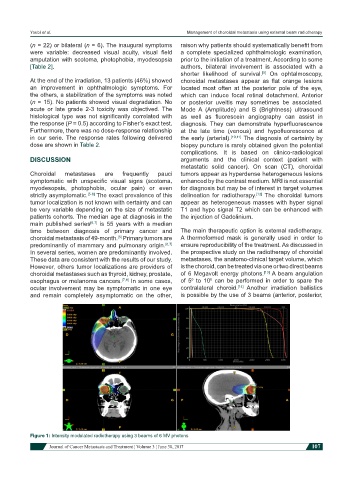
Figure 1: Intensity modulated radiotherapy using 3 beams of 6 MV photons
Journal of Cancer Metastasis and Treatment ¦ Volume 3 ¦ June 30, 2017 107