
Page 120 - Read Online
P. 120
Malik et al. LMS renal pelvis: a rare entity
scar, due to laproscopic cholecystectomy done 2 bed to a dose of 50 Gy/25#/5 weeks. Patient is doing
years back was visualized. No mass palpable. Triple well 1 year post treatment.
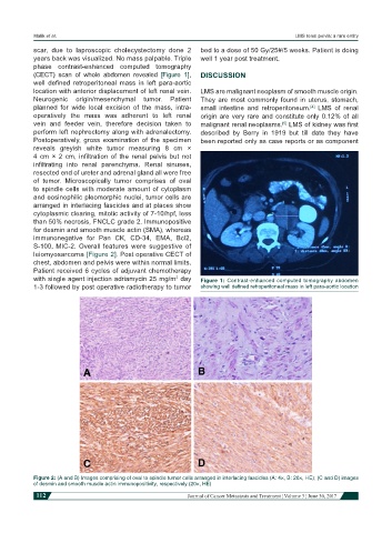
phase contrast-enhanced computed tomography
(CECT) scan of whole abdomen revealed [Figure 1], DISCUSSION
well defined retroperitoneal mass in left para-aortic
location with anterior displacement of left renal vein. LMS are malignant neoplasm of smooth muscle origin.
Neurogenic origin/mesenchymal tumor. Patient They are most commonly found in uterus, stomach,
planned for wide local excision of the mass, intra- small intestine and retroperitoneum. LMS of renal
[4]
operatively the mass was adherent to left renal origin are very rare and constitute only 0.12% of all
vein and feeder vein, therefore decision taken to malignant renal neoplasms. LMS of kidney was first
[5]
perform left nephrectomy along with adrenalectomy. described by Berry in 1919 but till date they have
Postoperatively, gross examination of the specimen been reported only as case reports or as component
reveals greyish white tumor measuring 8 cm ×
4 cm × 2 cm, infiltration of the renal pelvis but not
infiltrating into renal parenchyma. Renal sinuses,
resected end of ureter and adrenal gland all were free
of tumor. Microscopically tumor comprises of oval
to spindle cells with moderate amount of cytoplasm
and eosinophilic pleomorphic nuclei, tumor cells are
arranged in interlacing fascicles and at places show
cytoplasmic clearing, mitotic activity of 7-10/hpf, less
than 50% necrosis, FNCLC grade 2. Immunopositive
for desmin and smooth muscle actin (SMA), whereas
immunonegative for Pan CK, CD-34, EMA, Bcl2,
S-100, MIC-2. Overall features were suggestive of
leiomyosarcoma [Figure 2]. Post operative CECT of
chest, abdomen and pelvis were within normal limits.
Patient received 6 cycles of adjuvant chemotherapy
with single agent injection adriamycin 25 mg/m day Figure 1: Contrast-enhanced computed tomography abdomen
2
1-3 followed by post operative radiotherapy to tumor showing well defined retroperitoneal mass in left para-aortic location
Figure 2: (A and B) Images comprising of oval to spindle tumor cells arranged in interlacing fascicles (A: 4×, B: 20×, HE); (C and D) images
of desmin and smooth muscle actin immunopositivity, respectively (20×, HE)
112 Journal of Cancer Metastasis and Treatment ¦ Volume 3 ¦ June 30, 2017