
Page 121 - Read Online
P. 121
Malik et al. LMS renal pelvis: a rare entity
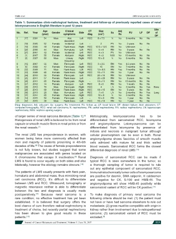
Table 1: Summarizes clinic-radiological features, treatment and follow-up of previously reported cases of renal
leiomyosarcoma in English literature in past 12 years
Site
Age Clinical CT Size Adj
No. Ref. Year Gender Side 2 Sx FU LF DF of
(year) symptoms diag (cm ) Rx
DF
1 [17] 2004 54 Male Abd Left RCC 8 × 7.5 N No 6 months No No
discomfort
2 [18] 2005 52 Female Incidental Left - 2 PN No 2 years No No
3 [19] 2006 48 Female Flank mass Right RCC 12.5 × 10.5 RN No Unknown - -
4 [20] 2006 44 Male Hematuria Left RCC 5 × 4 RN No 3 years No No
5 [21] 2007 42 Female Incidental Left RCC 5 × 3 PN No Unknown - -
6 [1] 2007 55 Female Abd pain Right RCC 4 × 2 NSS No 15 months No No
7 [8] 2007 60 Male Urinary Right RCC 10 × 8 N No 6 months No No
frequency
8 [15] 2007 55 Male Flank pain Left RCC 3 × 2.5 RN Yes 6.5 years No No
9 [6] 2009 42 Female Flank pain Right RCC 15 RN No 7 months No No
10 [22] 2009 71 Male Flank mass Left RCC 13 × 6.5 RN Yes 7 months No No
11 [23] 2009 65 Female Flank mass Right RCC 15 × 11 RN No 1 year No No
12 [24] 2010 55 Female Flank pain Left RCC 20 × 16 RN No Unknown - --
13 [25] 2011 57 Female Flank mass Left - 25 × 23 RN No 3 years No No
14 [26] 2011 65 Female Flank pain Right - 13 × 11 RN No 15 months No No
15 [27] 2012 74 Female Abd pain Left RCC 8 × 6 RN No 1 month No No
16 [28] 2013 70 Male Flank pain Right RCC 4 LN No 2 months No Yes Bone
17 [29] 2013 69 Female Flank mass Right RCC 18 × 15 RN No 5 years No No
18 [30] 2013 20 Male Hematuria Left RCC 7 × 6 LN No 2 months No Yes Lung
19 [31] 2014 65 Female Flank mass Right RCC 18 × 8 N No 2 months No No
20 [14] 2015 50 Female Flank pain Left - 10 × 6 RN No 1 year No No
21 [32] 2015 39 Male Flank mass Left RCC 16 × 14 RN No 1 year No No
Diag: diagnosis; Adj: adjuvant; Sx: surgery; Rx: treatment; FU: follow up; LF: local failure; DF: distant failure; Abd: abdomen; CT:
computed tomography; RCC: renal cell carcinoma; N: nephrectomy; PN: partial nephrectomy; RN: radical nephrectomy; LN: laparoscopic
nephrectomy; NSS: nephron sparing surgery
of larger series of renal sarcoma literature [Table 1]. Histologically, leiomyosarcoma has to be
[6]
Histogenesis of renal LMS is believed to be from renal differentiated from sarcomatoid RCC, leiomyoma
capsule or smooth muscle fibers in renal pelvis or from and angiomyolipoma. Leiomyosarcoma can be
the renal vessels. [7] differentiated from leiomyoma by presence of
mitosis and necrosis in malignant tumor although
The renal LMS has preponderance in women, with cellular pleomorphism can be seen in both. Renal
women being twice more commonly affected than angiomyolipoma shows fascicles of smooth muscle
men and majority of patients presenting in 4th-6th cells admixed with mature fat and thick walled
decades of life. The cause of female preponderance blood vessels. Sarcomatoid RCC forms the closest
[8]
is not fully known, but studies suggest that some differential diagnosis of renal LMS. [13]
malignancies are associated with genes located on
X chromosome that escape X inactivation. Renal Diagnosis of sarcomatoid RCC can be made if
[9]
LMS is found to occur equally on both sides and also typical RCC is seen somewhere in the tumor, so
bilaterally, however the etiology remains obscure. [10] a thorough sampling of tumor is required to rule
out any epithelial component of sarcomatoid RCC.
The patients of LMS usually presents with flank pain, Immunohistochemically tumor cells of leiomyosarcoma
hematuria and abdominal mass, thus mimicking renal are positive for desmin, SMA calponin, H caldesmon
cell carcinoma (RCC). It’s difficult to differentiate and negative for CK, S-100 and HMB-45. The
between LMS and RCC. Ultrasound, tomography or angiomyolipoma will show HMB-45 positivity while
magnetic resonance neither is able to differentiate sarcomatoid variant of RCC will be CK positive. [14]
between the two and diagnosis is usually made
postoperatively. Because renal sarcoma is a To make diagnosis of primary renal sarcoma the
[11]
rare condition; no effective treatment has yet been following criteria should be met: (1) the patient must
established. It is believed that surgery offers the not have or have had sarcoma elsewhere to rule out
best chance of cure therefore radical nephrectomy is metastasis; (2) gross must be compatible with origin in
treatment of choice, but recently partial nephrectomy kidney rather than involvement due to retroperitoneal
has been shown to give good results in these sarcoma; (3) sarcomatoid variant of RCC must be
patients. [12] excluded. [8]
Journal of Cancer Metastasis and Treatment ¦ Volume 3 ¦ June 30, 2017 113