
Page 63 - Read Online
P. 63
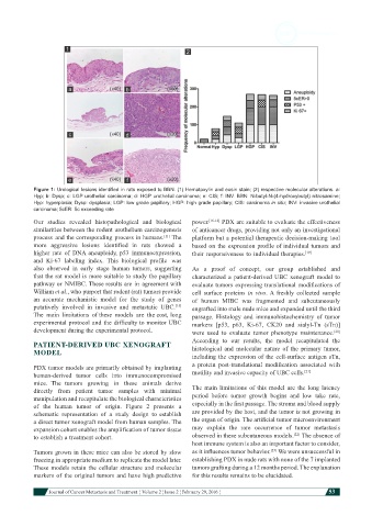
Figure 1: Urological lesions identified in rats exposed to BBN. (1) Hematoxylin and eosin stain; (2) respective molecular alterations. a:
            Hyp; b: Dysp; c: LGP urothelial carcinoma; d: HGP urothelial carcinoma; e: CIS; f: INV. BBN: N-butyl-N-(4-hydroxybutyl) nitrosamine;
            Hyp: hyperplasia; Dysp: dysplasia; LGP: low grade papillary; HGP: high grade papillary; CIS: carcinoma in situ; INV: invasive urothelial
            carcinoma; 5cER: 5c exceeding rate
            Our  studies  revealed  histopathological and  biological   power. [16-18]  PDX are suitable to evaluate the effectiveness
            similarities between the rodent urothelium carcinogenesis   of anticancer drugs, providing not only an investigational
            process  and the corresponding  process in humans.  The   platform but a potential therapeutic decision-making  tool
                                                     [11]
            more  aggressive  lesions  identified in  rats  showed  a   based on  the  expression profile of individual tumors and
            higher  rate  of  DNA aneuploidy,  p53  immunoexpression,   their responsiveness to individual therapies. [19]
            and  Ki-67  labeling  index.  This  biological  profile  was
            also  observed  in  early stage human  tumors,  suggesting   As a proof of concept, our group established and
            that the rat model is more suitable to study the papillary   characterized  a  patient-derived  UBC  xenograft  model  to
            pathway or  NMIBC. These results are  in  agreement with   evaluate tumors expressing translational modifications of
            William et al., who purport that rodent (rat) tumors provide   cell surface proteins  in vivo. A freshly collected sample
            an accurate mechanistic model  for  the  study  of  genes   of human MIBC was fragmented and subcutaneously
            putatively involved in  invasive and  metastatic  UBC.    engrafted into male nude mice and expanded until the third
                                                         [15]
            The  main limitations  of these models  are  the cost, long   passage.  Histology  and  immunohistochemistry  of  tumor
            experimental protocol and the  difficulty to monitor UBC   markers [p53, p63, Ki-67, CK20 and sialyl-Tn (sTn)]
            development during the experimental protocol.      were used to  evaluate  tumor phenotype maintenance.
                                                                                                            [20]
            PATIENT-DERIVED UBC XENOGRAFT                      According to our results, the model recapitulated the
            MODEL                                              histological and molecular nature of the primary tumor,
                                                               including the expression of the cell-surface antigen sTn,
            PDX tumor models are primarily obtained by implanting   a  protein  post-translational  modification  associated  with
            human-derived tumor  cells  into  immunocompromised   motility and invasive capacity of UBC cells. [21]
            mice.  The tumors  growing  in  these  animals derive
            directly from  patient  tumor  samples with  minimal   The main limitations of this model are the long latency
            manipulation and recapitulate the biological characteristics   period before tumor growth begins and low take rate,
            of  the  human tumor  of  origin. Figure 2  presents  a   especially in the first passage. The stroma and blood supply
            schematic  representation  of  a  study  design  to establish   are provided by the host, and the tumor is not growing in
            a direct tumor xenograft model from human samples. The   the organ of origin. The artificial tumor microenvironment
            expansion cohort enables the amplification of tumor tissue   may  explain  the  rare  occurrence  of  tumor  metastasis
                                                                                               [22]
            to establish a treatment cohort.                   observed in these subcutaneous models.  The absence of
                                                               host immune system is also an important factor to consider,
            Tumors grown in these mice can also be stored by slow   as it influences tumor behavior.  We were unsuccessful in
                                                                                        [23]
            freezing in appropriate medium to replicate the model later.   establishing PDX in nude rats with none of the 7 implanted
            These models retain the  cellular structure and molecular   tumors grafting during a 12 months period. The explanation
            markers  of  the  original tumors  and  have  high predictive   for this results remains to be elucidated.


                        Journal of Cancer Metastasis and Treatment  ¦ Volume 2 ¦ Issue 2 ¦ February 29, 2016 ¦  53