
Page 266 - Read Online
P. 266
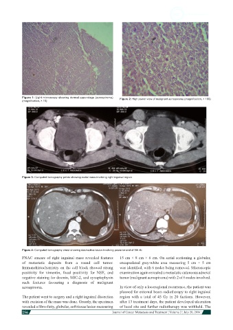
Figure 1: Light microscopy showing dermal appendage (acrospiroma)   Figure 2: High power view of malignant acrospiroma (magnification, × 100)
            (magnification, × 10)


            Figure 3: Computed tomography pelvis showing nodal mass involving right inguinal region


            Figure 4: Computed tomography chest showing destructive lesion involving posterior end of 9th rib
            FNAC smears of right inguinal mass revealed features   15 cm × 8 cm × 6 cm. On serial sectioning a globular,
            of metastatic deposits from a round cell tumor.   encapsulated  grey-white  area  measuring  5 cm × 5 cm
            Immunohistochemistry on the cell block showed strong   was identified, with 6 nodes being removed. Microscopic
            positivity for vimentin, focal positivity for NSE, and   examination again revealed a metastatic cutaneous adnexal
            negative staining for desmin, MIC-2, and synaptophysin   tumor (malignant acrospiroma) with 2 of 6 nodes involved.
            such features favouring a diagnosis of malignant
            acrospiroma.                                      In view of only a locoregional recurrence, the patient was
                                                              planned for external beam radiotherapy to right inguinal
            The patient went to surgery and a right inguinal dissection   region with a total of 45 Gy in 20 factions. However,
            with excision of the mass was done. Grossly, the specimen   after 13 treatment days, the patient developed ulceration
            revealed a fibro-fatty, globular, soft tissue lesion measuring   of local site and further radiotherapy was withheld. The
            256
                                                                                                                        Journal of Cancer Metastasis and Treatment ¦ Volume 2 ¦ July 20, 2016 ¦