
Page 150 - Read Online
P. 150
the newer techniques that conform to the optimum dose of
the CTV or the shape of the target volume. Even a little
geometrical movement could result in an underdosing to the
target volume or conversely, delivering high undesirable
doses to the surrounding normal tissues. These effects
highlight the importance of accurate margin determination.
This pilot study was conducted to define the daily uterine
shift in patients undergoing external radiotherapy on linear
accelerators with IMRT technique using IGRT with the
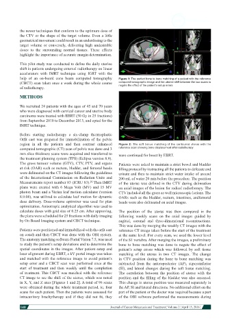
help of an on-board cone beam computed tomography Figure 1: The perfect bone to bone matching of a patient with the reference
(CBCT) scan taken once a week during the whole course computed tomographic image and the uterine shift between the two scans to
negate the effect of the patient’s setup errors
of radiotherapy.
METHODS
We recruited 24 patients with the ages of 45 and 70 years
who were diagnosed with cervical cancer and uterine body
carcinoma were treated with EBRT (50 Gy in 25 fractions)
from September 2010 to December 2013, and opted for the
IMRT technique.
Before starting radiotherapy a six-clamp thermoplastic
Orfit cast was prepared for immobilization of the pelvic
region in all the patients and then contrast enhanced Figure 2: The soft tissue matching of the contoured uterus with the
computed tomographic (CT) scan of pelvis was done and 3 reference scan showing bone displacement after radiotherapy
mm slice thickness scans were acquired and transferred to were continued for boost by EBRT.
the treatment planning system (TPS) (Eclipse version 8.9).
The gross tumour volume (GTV), CTV, PTV, and organs Patients were asked to maintain a strict bowel and bladder
at risk (OAR) such as rectum, bladder, and femoral heads filling protocol by instructing all the patients to defecate and
were delineated on the CT images following the guidelines urinate and then to maintain strict water intake of around
of the International Commission on Radiation Units and 200 mL of water 20 min before the procedure. The position
Measurements report number 83 (ICRU 83). Then IMRT of the uterus was defined in the CTV during delineation
[4]
plans were created with 6 Mega Volt (MV) and 15 MV on axial images of the lesion for radical radiotherapy. The
photon beam and a Varian leaf motion calculator (version CTV included all the gross as well microscopic lesions. The
8.9.08), was utilized to calculate leaf motion for dynamic OARs such as the bladder, rectum, intestines, andfemoral
dose delivery. Dose-volume optimizer was used for plan heads were also delineated on axial images.
optimization. Anisotropic analytical algorithm was used to
calculate doses with grid size of 0.25 cm. After approving, The position of the uterus was then compared in the
the plans were scheduled for 25 fractions with daily imaging following weekly scans on the axial images guided by
by On Board Imaging system and CBCT technique. sagittal, coronal and three-dimensional reconstructions.
This was done by merging the weekly CT images with the
Patients were positioned and immobilized with the orfit cast reference CT image taken before the start of the treatment
on couch and then CBCT was done with the OBI system. at the same level. For every scan, we used the lower level
The anatomy matching software Portal Vision 7.5, was used of the S1 vertebra. After merging the images, a preliminary
to study the patient’s setup deviations and to determine the bone to bone matching was done to negate the effect of
spatial coordinates in the images. After patient setup and patient’s setup errors which was followed by soft tissue
laser alignment during EBRT, a kV portal image was taken matching of the uterus in two CT images. The change
and matched with the reference image to avoid patient’s in CTV position during the bone to bone matching was
setup error and a CBCT scan was performed once at the subtracted from the anteroposterior (AP), superoinferior
start of treatment and then weekly until the completion (SI), and lateral changes during the soft tissue matching.
of treatment. This CBCT was matched with the reference The correlation between the position of uterus with the
CT image to see the shift of the uterus, which was noted position and the filling of the bladder was also assessed.
in X, Y, and Z axes [Figures 1 and 2]. A total of 96 scans This change in uterus position was measured separately in
were obtained during the whole treatment period, ie, four the AP, SI and lateral directions. No additional effort on the
scans for each patient. Then the patients were assessed for part of the patient or the doctor was required because a part
intracavitory brachytherapy and if they did not fit, they of the OBI software performed the measurements during
140
Journal of Cancer Metastasis and Treatment ¦ Volume 2 ¦ April 15, 2016 ¦