
Page 152 - Read Online
P. 152
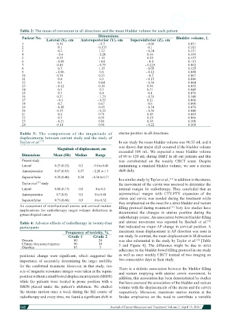
Table 2: The mean of movement in all directions and the mean bladder volume for each patient
                                                    Dimensions
            Patient No.  Lateral (X), cm  Anteroposterior (Y), cm   Superoinferior (Z), cm  Bladder volume, L
            1                 0.16                  - 0.3                   - 0.06                0.031
            2                  0.1                 - 0.133                   0.1                  0.111
            3                  0.2                  1.32                    - 0.24                0.151
            4                 - 0.6                - 2.28                    0.16                 0.189
            5                 0.25                  1.15                     0.55                 0.157
            6                 - 0.08                1.04                    - 0.4                 0.113
            7                 0.45                 - 0.75                   - 0.225               0.062
            8                  0.3                  1.15                    - 0.05                0.125
            9                 - 0.06                0.6                     - 0.12                0.088
            10                0.36                  0.55                    - 0.2                 0.067
            11                 0.4                  0.3                     - 0.15                0.046
            12                 0.1                  0.04                    - 0.36                0.064
            13                - 0.12                0.18                     0.56                 0.055
            14                 0.1                  0.3                      0.71                 0.049
            15                 0.3                  0.4                      0.4                  0.076
            16                0.31                 - 1.21                   - 0.35                0.140
            17                - 0.1                - 0.53                    0.21                 0.096
            18                 0.2                  0.67                     0.6                  0.090
            19                - 0.45                0.45                    - 0.34                0.070
            20                0.15                 - 0.22                   - 0.56                0.040
            21                 0.2                  0.71                     0.43                 0.083
            22                 0.3                  0.51                     0.25                 0.066
            23                - 0.21                0.81                     0.16                 0.100
            24                0.14                  0.91                    - 0.22                0.105

           Table 3:  The comparison of the magnitude of       uterine position in all directions.
           displacements between current study and the study of
           Taylor et al. [11]                                 In our study the mean bladder volume was 90.55 mL and it
                              Magnitude of displacement, cm   was shown that major shift occurred if the bladder volume
                                                              exceeded  100 mL.  We expected a mean bladder volume
            Dimensions      Mean (SD)  Median     Range       of 80 to 120 mL during IMRT in all our patients and this
            Present study                                     was  corroborated on the weekly CBCT scans. Despite
            Lateral          0.23 (0.22)  0.2    - 0.6 to 0.45  maintaining a standard bladder volume, we saw a uterine
            Anteroposterior  0.67 (0.83)  0.57   - 2.28 to 1.3  shift daily.
            Superoinferior   0.29 (0.40)  0.24  - 0.36 to 0.71  In a similar study by Taylor et al.,  in addition to the uterus,
                                                                                        [11]
                    [11]
            Taylor et al.  study                              the movement of the cervix was assessed to determine the
            Lateral          0.08 (0.13)  0.0     0 to 0.5    internal  margin for radiotherapy. They concluded  that  an
            Anteroposterior   0.7 (0.9)   0.5     0 to 0.48   asymmetrical  margin with CTV-PTV expansion of the
            Superoinferior   0.71 (0.68)  0.5     0 to 0.32   uterus and cervix was needed during the treatment while
           An assessment of interfractional uterine and cervical motion:   they emphasized on the need for a strict bladder and rectum
                                                              filling protocol during treatment.  Very few studies have
                                                                                         [11]
           implications for radiotherapy target volume definition in
           gynaecological cancer                              documented  the changes in uterine  position  during the
                                                              radiotherapy course. An association between bladder filling
           Table 4: Adverse effects of radiotherapy in twenty-four   and uterine  movement  was reported  by Buchali  et  al. [12]
           participants                                       that indicated no major AP change in cervical position. A
                                   Frequency of toxicity, %   maximum mean displacement in AP direction was seen in
                                    Grade 1      Grade 2      our study. In contrast, the mean displacement in SI direction
            Dysuria                    80           20        was also substantial in the study by Taylor et al.  [Table
                                                                                                      [11]
            Urinary frequency/urgency  90           10        3 and Figure 4].  The difference might be due to strict
            Diarrhea                   95           5
                                                              adherence to the bladder-bowel filling protocol in our study
           positional  change  were  significant,  which  suggested  the   as well as once weekly CBCT instead of two imaging on
           importance  of  accurately  determining  the  target  mobility   two consecutive days in their study.
           for the conformal treatment. However, in that study, two   There is a definite association between the bladder filling
           sets of magnetic resonance images were taken in the supine   and  rectum  emptying  with  uterine  cervix  movement.  In
           position without a small bowel displacement system (SBDS)   addition, this association has been demonstrated by studies
           while the patients were treated  in prone position with a   that have assessed the association of the bladder and rectum
           SBDS  placed  under  the  patient’s  abdomen.  We  studied   volume with the displacements of the uterus and the cervix
           the uterine motion once a week during the full course of   respectively. Moreover, maximum uterine  motion  at the
           radiotherapy and every time, we found a significant shift in   fundus emphasizes  on the  need  to  contribute  a  variable

           142
                                                                                                                        Journal of Cancer Metastasis and Treatment ¦ Volume 2 ¦ April 15, 2016 ¦