
Page 77 - Read Online
P. 77
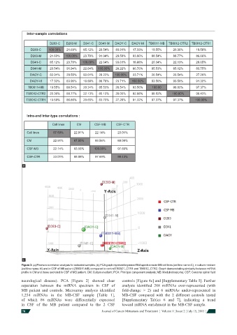
Inter-sample correlations


                          D283-C  D283-M   D341-C  D341-M  DAOY-C  DAOY-M   TB0011-MB  TB0012-CTR2  TB0012-CTR1
                D283-C    100.00%  21.69%  85.12%  20.54%  83.91%   17.33%   19.55%     20.36%      19.58%
                D283-M    21.69%  100.00%  23.70%  91.94%  29.50%   83.96%   88.54%     88.77%      86.66%
                D341-C    85.12%  23.70%  100.00%  22.64%  93.61%   18.88%   20.34%     22.13%      20.65%
                D341-M    20.54%  91.94%   22.64%  100.00%  28.22%  86.76%   85.53%     85.12%      83.75%
                DAOY-C    83.91%  29.50%   93.61%  28.22%  100.00%  23.71%   26.54%     26.54%      27.26%
                DAOY-M    17.33%  83.96%   18.88%  86.76%  23.71%  100.00%   93.56%     93.56%      91.32%
               TB0011-MB  19.55%  88.54%   20.34%  85.53%  26.54%   93.56%   100.00     98.02%      97.37%
              TB0012-CTR2  20.36%  88.77%  22.13%  85.12%  28.05%   92.88%   98.02%     100.00%     98.43%
              TB0012-CTR1  19.58%  86.66%  20.65%  83.75%  27.26%   91.32%   97.37%     97.37%     100.00%


              Intra-and inter-type correlations :

                           Cell lines   CM      CSF-MB     CSF-CTR

               Cell lines   87.59%    22.91%     22.14%     23.01%
               CM           22.91%    87.55%     93.56%     88.08%

               CSF-MB       22.14%    93.56%    100.00%     97.69%

               CSF-CTR      23.01%    88.08%     97.69%     98.43%
            a


            b
            Figure 3: (a) Pearson correlation analysis for indicated samples; (b) PCA graph representing microRNA spectra inside MB cell lines (cell line name-C), in culture medium
            (cell line name-M) and in CSF of MB patient (TB0011-MB) compared to control TB0021_CTR1 and TB0012_CTR2. Graph demonstrating similarity between miRNA
            profi le in CM and those secreted in CSF of MB patient. CM: Culture medium; PCA: Principal component analysis; MB: Medulloblastoma; CSF: Cerebral spinal fl uid

            neurological disease). PCA [Figure 2] showed clear   controls [Figure 4c] and [Supplementary Table 5]. Further
            separation between the miRNA spectrum in CSF of   analysis identifi ed 268 miRNAs over-represented (with
            MB patient and controls. Microarray analysis identifi ed   fold-change > 2) and 6 miRNAs under-represented in
            1,254 miRNAs in the MB-CSF sample [Table 1],      MB-CSF compared with the 2 different controls tested
            of which 86 miRNAs were differentially expressed   [Supplementary  Tables 6 and 7], indicating a trend
            in CSF of the MB patient compared to the 2 CSF    toward miRNA enrichment in the MB-CSF sample.
            70                                      Journal of Cancer Metastasis and Treatment  ¦  Volume 1 ¦ Issue 2 ¦ July 15, 2015 ¦