
Page 35 - Read Online
P. 35
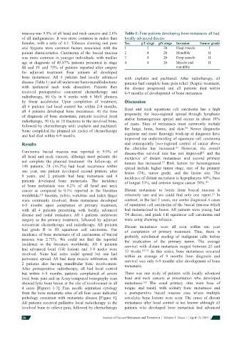
mucosa was 9.9% of all head and neck cancers and 2.6%   Table 1: Four patients developing bone metastases all had
            of all malignancies. It was more common in males than   locally advanced disease
            females, with a ratio of 4:1. Tobacco chewing and poor   No.  pT stage  pN stage  Invasion  Tumor grade
            oral hygiene were common factors associated with the   1     2       2b     Deep muscle     I
            patient characteristics. Carcinoma of the buccal mucosa   2  4       2b     Mandible        II
            was more common in younger individuals, with median   3      4       2b     Deep muscle     II
            age at diagnosis of 45.87% patients presented in stage   4   4       2b     Muscle and      II
            III and IV and 75% of patients reported after surgery                       mandible
            for adjuvant treatment. Four patients all developed
            bone metastases.    All 4 patients had locally advanced   with cisplatin and paclitaxel.  After radiotherapy, all
            disease [Table 1] and all underwent hemi-mandibulectomy   patients had complete bone pain relief. Despite treatment,
            with ipsilateral neck node dissection. Patients then   the disease progressed and all patients died within
            received postoperative concurrent chemotherapy and   6-9 months of development of bone metastasis.
            radiotherapy, 60 Gy in 6 weeks with 6 MeV photons
            by linear accelerator. Upon completion of treatment,   Discussion
            all 4 patients had local control but within 2-8 months,
            all 4 patients developed bone metastases.  At the time   Head and neck squamous cell carcinoma has a high
            of diagnosis of bone metastasis, patients received local   propensity for loco-regional spread through lymphatic
            radiotherapy, 30 Gy in 10 fractions to the involved bone,   and/or hematogenous spread and occurs in about 10%
            followed by chemotherapy with cisplatin and paclitaxel.   of cases. Sites of metastases most commonly include
                                                                                            [4]
            None completed the planned six cycles of chemotherapy   the lungs, brain, bones, and skin.  Newer diagnostic
            and had died within 6-9 months.                   regimens and more thorough work-up at diagnosis have
                                                              improved our understanding of squamous cell carcinoma
            Results                                           and consequently loco-regional control of cancer above
                                                                                       [5]
                                                              the clavicles has increased.  However, the overall
            Carcinoma buccal mucosa was reported in 9.9% of   disease-free survival rate has not improved  and the
                                                                                                    [6]
            all head and neck cancers, although most patients did   incidence of distant metastases and second primary
            not complete the planned treatment. On follow-up, of   tumors has increased.  Risk factors for hematogenous
                                                                                 [7]
            148 patients, 24 (16.2%) had local recurrence within   spread include higher tumor stage, size of the primary
            one year, one patient developed second primary after   lesion (T4), tumor grade, and the lesion site.  The
            8 years, and 2 patients had lung metastasis and 4   incidence of distant metastasis is hypopharynx 60%, base
            patients developed bone metastasis.  The incidence   of tongue 53%, and anterior tongue cancer 50%. [8]
            of bone metastasis was 0.2% of all head and neck
            cancer as compared to 0.1% reported in the literature   Distant metastasis to bones from buccal mucosa is
                                                                                                         [9]
            worldwide.  Sacrum, pelvis, vertebrae and index  fi nger   extremely rare and we could  fi nd only one report.  In
                     [1]
            were commonly involved. Bone metastases developed   contrast, in the last 5 years, our center diagnosed 4 cases
            6-9 months upon completion of primary treatment,   of squamous cell carcinoma of the buccal mucosa which
            with all 4 patients presenting with locally advanced   had metastasized to bones. All patients were young, had
            disease and nodal metastasis.  All 4 patients underwent   T4 disease, and grade I-II squamous cell carcinoma and
            surgery as the primary treatment, followed by adjuvant   were using chewing tobacco.
            concurrent chemotherapy and radiotherapy.  All patients   Distant metastases were all seen within one year
            had grade II to III squamous cell carcinoma. Our   of completion of primary treatment.  Thus, there is
            incidence of bone metastasis of all carcinomas of buccal   probably subclinical seeding of malignant cells before
            mucosa was 2.71%.  We could not  fi nd the reported   the eradication of the primary tumor.  The average
            incidence in the literature worldwide.  All 4 patients   survival with distant metastasis ranged between 21 and
            had advanced local (T4) lesions and 3-8 nodes were   33 weeks. [10,11]  In this series, bone metastases occurred
            involved. None had extra nodal spread but one had   within an average of 9 months from diagnosis and
            perineural spread. All had deep muscle infi ltration,  with   survival was only 6-9 months after development of bone
            2 patients also having mandibular bone involvement.   metastasis.
            After postoperative radiotherapy, all had local control
            but within 6-9 months, patients complained of severe   There was one study of patients with locally advanced
            local bone pain and   an   X-ray/computed tomography scan   head and neck cancers at presentation who developed
                                                                       [12]
            showed lytic bone lesion at the site of involvement in all   metastases.   The usual primary sites were base of
            4 cases [Figures 1-3]. Fine needle aspiration cytology   tongue and tonsil, with solitary bone metastases and
            from the bone metastatic sites of all four cases indicated   a postoperative buccal mucosa case where multiple
            pathology consistent with metastatic disease [Figure 4].   osteolytic bone lesions were seen.  The cause of distant
            All patients received palliative local radiotherapy to the   metastases after local control is not known although all
            involved bone to relieve pain, followed by chemotherapy   patients who developed bone metastasis had advanced

            28                                      Journal of Cancer Metastasis and Treatment  ¦  Volume 1 ¦ Issue 1 ¦ April 15, 2015 ¦