
Page 36 - Read Online
P. 36
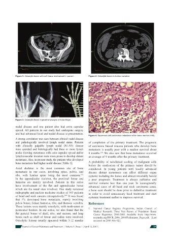
Figure 1: Osteolytic lesion with soft tissue involvement in sacrum  Figure 2: Osteolytic lesion in lumber vertebra


            Figure 3: Osteolytic lesion in proximal phalynx of index fi nger

            nodal disease and one patient also had extra capsular
            spread. All patients in our study had undergone surgery
            and had advanced local and nodal disease at presentation.
                                                              Figure 4: Squamous cell carcinoma metastasis within bone marrow (×40)
                A strong correlation was seen between clinical nodal disease
            and pathologically involved lymph nodal status. Patients   of completion of the primary treatment.  The prognosis
            with clinically palpable lymph nodal (N1-N3) disease   of carcinoma buccal mucosa patients who develop bone
            were operated and histologically had three or more lymph   metastasis is usually poor with a median survival about
            nodes showing metastases with extra capsular spread and/or   8 months.  We also saw that bone metastases occurred
                                                                      [15]
            lymphovascular invasion were more prone to develop distant   an average of 9 months after the primary treatment.
            metastasis. Also, in present study, the patients who developed
            bone metastasis had higher nodal disease [Table 1].  A probability of subclinical seeding of malignant cells
                                                              before the eradication of the primary tumor should be
                Axial skeleton is the most common site of bone   considered. In young patients with locally advanced
            metastasis in our cases, involving spine, pelvis, and   disease distant metastases can affect different organ
            ribs, with lumbar spine being the most common.    systems including the bones and almost invariably herald
                                                         [13]
            In the appendicular skeleton, the proximal femur and   a poor prognosis.  Treatment is always palliative and
            humerus are mainly involved. Patients in this series   survival remains less than one year. In locoregionally
            have involvement of the  fl at and appendicular bones   advanced cases of all head and neck carcinoma cases,
            which are the usual sites involved. One study reviewed   a bone scan should be done prior to defi nitive treatment
            radiographs and nuclear medicine studies of 363 patients   in order to avoid unnecessary local treatment and start
            of head and neck cancers retrospectively.  It was found   systemic treatment earlier to improve survival.
                                              [14]
            that 1% developed bone metastasis, mainly involving
            pelvic bones, femur, humerus, ribs, and thoracic vertebra.   References
            These lesions were mainly osteolytic, with moth-eaten or
            permeated borders. In our series, we also found that the     1.   National Cancer Registry Programme, Indian Council of
                                                                  Medical Research.  Three  Year Report of Population Based
            fl at parietal bones of skull, ribs, and sacrum, and long   Cancer Registries; 2006-2008.  Available from: http://www.
            bones such as shaft of femur and radius were involved.   ncrpindia.org/PBCR_2006_2008/Preliminary_Pages.pdf. [Last
            Osteolytic lesions usually appeared within 3-12 months   accessed on 2010 Nov 02].

                Journal of Cancer Metastasis and Treatment  ¦  Volume 1 ¦ Issue 1 ¦ April 15, 2015 ¦       29