
Page 30 - Read Online
P. 30
express HER2 at all. FISH analysis was conducted in the data revealed no HER2 amplifi cation in these cases.
those cases with the HER2 score of 2+ or more and Among 1+ cases, FISH was carried out in only two
selected carcinomas showing high grade, high Ki-67
Table 1: Clinicopathological and HER2 concordance in value, N+ status, and the absence of endocrine receptors
62 GC patients expression, but no HER2 amplifi cation was identifi ed.
Discordant GC Concordant GC P HER2 was amplifi ed in 14 BC cases (21.54%) but there
Gender was no HER2 amplifi cation in these 51 cases (78.46%).
Male 4 36 0.739 The overall concordance rate was 95.39%, whereas
Female 2 20 changes in HER2 status between primary carcinoma and
Site corresponding synchronous metastases were evidenced
Lower 3 27 0.389 in 3 (4.61%) cases [Table 3]. Two of the discordant
Middle 1 21 cases were HER2 negative in the primitive tumor
Upper 2 8 but positive in the metastasized tumors [Figure 2a
Lauren histotype and b], whereas one case was HER2 positive in the
Intestinal 3 31 0.369 primary BC and turned to negative in the metastatic
Diffuse 1 19 tumor [Figure 2c and d and Table 4].
Mixed 2 6 After that, we performed statistical analyses and found
WHO histotype that the K value for the concordance rate in the HER2
Tubular 4 31 0.672 status between primitive tumors and metastases was
Poorly cohesive 1 19 0.651 (substantial agreement). HER2 amplifi cation was
Mixed 1 6
Grade signifi cantly more frequent in the intestinal-type GC than
that of diffuse-type while no signifi cant differences in
Low 4 28 0.728 HER2 expression were noted among BC histology types.
High 2 28
Stage No statistical signifi cant correlation emerged between
I-II 3 21 0.875 HER2 and clinicopathological parameters (hormone
III-IV 3 35 receptors, growth fraction, pT, pN, and grade) either in
T GC as well as BC.
1-2 2 18 0.689 Discussion
3-4 4 38
N In the current study, we retrospectively analyzed
1 3 24 0.922 HER2 expression in surgical GC and BC specimens
2-3 3 32 versus the corresponding metastatic lymph nodes.
Our results fi rstly confi rmed the presence of a high
GC: Gastric carcinoma; HER2: Human epidermal growth
factor receptor 2 level of concordance in HER2 status between the
primary GC/BC and their corresponding lymph node
a b
a b
c d
c d
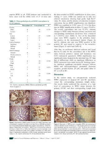
Figure 1: Expression of HER2 protein. A score of 3+ HER2 expression was
encountered in neoplastic elements in a primary GC (a, ×200) but vanished in Figure 2: Expression of HER2 protein. A negative HER2 immunostaining in
the corresponding metastatic lymph node (negative conversion) (b, ×160) (IHC, a primary infi ltrative ductal carcinoma (a, ×160) became positive in the lymph
Mayer’s hematoxylin counterstain). HER2 immunohistochemical negative node metastasis (positive conversion) (b, ×120). The strong and complete
staining in primary GC (c, ×200), demonstrated a positive reactivity in the HER2 immunoreactivity in a case of primary BC (c, ×200) was not present in
metastatic synchronous lymph node (positive conversion) (d, ×200) (IHC, the synchronous lymph nodal metastasis (negative conversion) (d, ×200) (IHC,
Mayer’s hematoxylin counterstain). HER2: Human epidermal growth factor Mayer’s hematoxylin counterstain). HER2: Human epidermal growth factor
receptor 2; GC: Gastric carcinomas; IHC: Immunohistochemistry receptor 2; BC: Breast carcinoma; IHC: Immunohistochemistry
Journal of Cancer Metastasis and Treatment ¦ Volume 1 ¦ Issue 1 ¦ April 15, 2015 ¦ 23