
Page 101 - Read Online
P. 101
Page 4 of 11 Jalal et al. J Cancer Metastasis Treat 2023;9:11 https://dx.doi.org/10.20517/2394-4722.2022.122
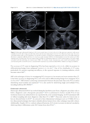
Figure 1. Endoscopic ultrasound imaging of (A) A microcystic lesion (circle) in the tail of the pancreas consistent with serous
cystadenoma (SCA). (B) A large uniocular thin-walled cyst (arrows) measuring 33 mm × 27 mm in the body of pancreas. The cyst is
uniformly anechoic with no solid component. The cyst has no evident communication with the pancreatic duct on the static image and
therefore is consistent with the definition of a mucinous cystic neoplasm (MCN). (C) Multiseptated (arrow) thin-walled cyst in the
head and neck of the pancreas. The cyst communicates with the non-dilated pancreatic duct, but this is not evident on the static image,
consistent with the definition of a branch-duct IPMN. (D) Another cluster of grape-like cysts (small multiseptated cysts)
communicating with a dilated PD (arrow), consistent with mixed main-duct and branch-duct IPMNs. Patient is on images surveillance.
The accuracy of CT scans in diagnosing PCLs has been reported as 39%-61.4%, while its accuracy in
[27]
differentiating benign from malignant lesions is 61.9%-80% . One of the drawbacks of CT scans,
particularly for patients requiring surveillance, is the repeated exposure to ionizing radiation, which
[28]
increases cancer risk .
MRI is the technique of choice for investigating PCLs because it is less invasive and more sensitive than CT.
It has better accuracy in diagnosing PCLs (50%-80%) and in differentiating benign from malignant PCLs
(55.6%-87%) . MRI is better at detecting communication between the pancreatic duct and a PCL as well as
[27]
identifying mural nodules and septations . It is also useful for identifying single and multiple PCLs,
[14]
including multifocal BD-IPMNs .
[28]
Endoscopic ultrasound
Endoscopic ultrasound (EUS) has evolved during the last thirty years from a diagnostic procedure only to
both a diagnostic and a therapeutic procedure . EUS is performed to further stratify PCLs if any
[29]
worrisome features (as outlined above) are reported in the cross-sectional imaging. An EUS assessment with
or without sampling adds more diagnostic value than CT or MRI . Pancreatic cysts of 20 mm in size or
[26]
less are detected better by EUS . In contrast to three-dimensional techniques such as CT/MRI, EUS is a
[30]
two-dimensional technique that helps to identify adverse features those other techniques may fail to detect.
Additionally, Doppler EUS or contrast-enhanced harmonic EUS reveals increases in internal vascularity,
[31]
which may help to differentiate cystic NET lesions from other PCLs .