
Page 22 - Read Online
P. 22
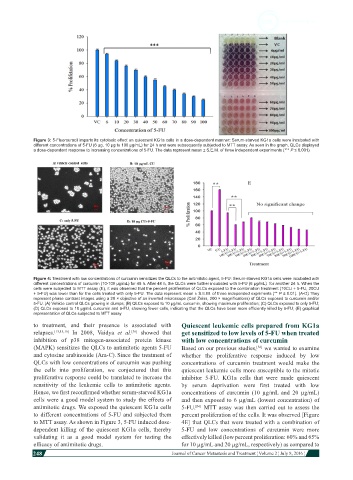
Figure 3: 5-Fluorouracil imparts its cytotoxic effect on quiescent KG1a cells in a dose-dependent manner: Serum-starved KG1a cells were incubated with
            different concentrations of 5-FU (6 µg, 10 µg to 100 µg/mL) for 24 h and were subsequently subjected to MTT assay. As seen in the graph, QLCs displayed
            a dose-dependent response to increasing concentrations of 5-FU. The data represent mean ± S.E.M. of three independent experiments (*** P ≤ 0.001)


            Figure 4: Treatment with low concentrations of curcumin sensitizes the QLCs to the antimitotic agent, 5-FU: Serum-starved KG1a cells were incubated with
            different concentrations of curcumin (10-100 µg/mL) for 48 h. After 48 h, the QLCs were further incubated with 5-FU (6 µg/mL)  for another 24 h. When the
            cells were subjected to MTT assay (E), it was observed that the percent proliferation of QLCs exposed to the combination treatment (10CU + 5-FU, 20CU
            + 5-FU) was lower than for the cells treated with only 5-FU. The data represent mean ± S.E.M. of three independed expriments (** P ≤ 0.01). (A-D) They
            represent phase contrast images using a 20 × objective of an inverted microscope (Carl Zeiss, 200 × magnifications) of QLCs exposed to curcumin and/or
            5-FU. (A) Vehicle control QLCs growing in clumps; (B) QLCs exposed to 10 µg/mL curcumin, showing maximum proliferation; (C) QLCs exposed to only 5-FU;
            (D) QLCs exposed to 10 µg/mL curcumin and 5-FU, showing fewer cells, indicating that the QLCs have been more efficiently killed by 5-FU; (E) graphical
            representation of QLCs subjected to MTT assay

            to treatment, and their presence is associated with   Quiescent leukemic cells prepared from KG1a
            relapses. [13,15,16]  In 2008,  Vaidya  et al.  showed that   get sensitized to low levels of 5-FU when treated
                                             [36]
            inhibition of p38 mitogen-associated protein kinase   with low concentrations of curcumin
            (MAPK) sensitizes the QLCs to antimitotic agents 5-FU   Based on our previous studies,  we wanted to examine
                                                                                        [36]
            and cytosine arabinoside (Ara-C). Since the treatment of   whether  the  proliferative  response  induced by  low

            QLCs with low concentrations of curcumin was pushing   concentrations of curcumin treatment would make the
            the cells into proliferation, we conjectured that this   quiescent leukemic cells more susceptible to the mitotic
            proliferative response could be translated to increase the   inhibitor 5-FU. KG1a cells that were made quiescent
            sensitivity of the leukemic cells to antimitotic agents.   by  serum  deprivation  were  first  treated  with  low
            Hence, we first reconfirmed whether serum-starved KG1a   concentrations of curcumin (10 µg/mL and 20 µg/mL)
            cells were a good model system to study the effects of   and then exposed to 6 µg/mL (lowest concentration) of
            antimitotic drugs. We exposed the quiescent KG1a cells   5-FU.   MTT  assay  was  then  carried  out  to  assess  the
                                                                   [36]
            to different concentrations of 5-FU and subjected them   percent proliferation of the cells. It was observed [Figure
            to MTT assay. As shown in Figure 3, 5-FU induced dose-  4E] that QLCs that were treated with a combination of
            dependent killing of the quiescent KG1a  cells, thereby   5-FU and low concentrations  of curcumin were more
            validating it as a good model system for testing the   effectively killed (low percent proliferation: 60% and 65%
            efficacy of antimitotic drugs.                    for 10 µg/mL and 20 µg/mL, respectively) as compared to
            248
                                                                                                                          Journal of Cancer Metastasis and Treatment ¦ Volume 2 ¦ July 8, 2016 ¦