
Page 23 - Read Online
P. 23
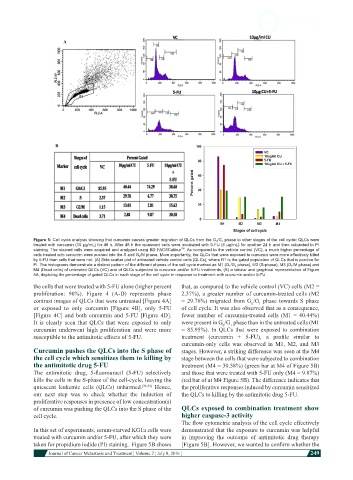
Figure 5: Cell cycle analysis showing that curcumin causes greater migration of QLCs from the G 0 /G 1  phase to other stages of the cell cycle: QLCs were
            treated with curcumin (10 µg/mL) for 48 h. After 48 h the quiescent cells were incubated with 5-FU (6 µg/mL) for another 24 h and then subjected to PI
                                                              TM
            staining. The stained cells were acquired and analyzed using BD FACSCalibur . As compared to the vehicle control (VC), a much higher percentage of
            cells treated with curcumin were pushed into the S and G 2 /M phase. More importantly, the QLCs that were exposed to curcumin were more effectively killed
            by 5-FU than cells that were not. (A) Side scatter plot of untreated vehicle control cells (QLCs), where R1 is the gated population of QLCs that is positive for
            PI. The histograms demonstrate a distinct pattern of the different phases of the cell cycle marked as M1 (G 1 /G 0  phase), M2 (S-phase), M3 (G 2 /M phase) and
            M4 (Dead cells) of untreated QLCs (VC) and of QLCs subjected to curcumin and/or 5-FU treatments; (B) a tabular and graphical representation of Figure
            5A, depicting the percentage of gated QLCs in each stage of the cell cycle in response to treatment with cucrumin and/or 5-FU
            the cells that were treated with 5-FU alone (higher percent   that, as compared to the vehicle control (VC) cells (M2 =
            proliferation: 90%). Figure 4 (A-D) represents phase   2.37%), a greater number of curcumin-treated cells (M2
            contrast images of QLCs that were untreated [Figure 4A]   = 29.76%) migrated from G /G  phase towards S phase
                                                                                        1
                                                                                      0
            or exposed to only curcumin [Figure 4B], only 5-FU   of cell cycle. It was also observed that as a consequence,
            [Figure 4C] and both curcumin and 5-FU [Figure 4D].   fewer number of curcumin-treated cells (M1 = 40.44%)
            It is clearly seen that QLCs that were exposed to only   were present in G /G  phase than in the untreated cells (M1
                                                                               1
                                                                            0
            curcumin underwent high proliferation and were more   = 85.95%). In QLCs that  were exposed to combination
            susceptible to the antimitotic effects of 5-FU.   treatment  (curcumin  +  5-FU),  a  profile  similar  to
                                                              curcumin-only  cells  was observed in M1, M2, and M3
            Curcumin pushes the QLCs into the S phase of      stages. However, a striking difference was seen at the M4
            the cell cycle which sensitizes them to killing by   stage between the cells that were subjected to combination
            the antimitotic drug 5-FU                         treatment (M4 = 30.58%) (green bar at M4 of Figure 5B)
            The  antimitotic  drug,  5-fluorouracil  (5-FU)  selectively   and those that were treated with 5-FU only (M4 = 9.87%)
            kills the cells in the S-phase of the cell-cycle, leaving the   (red bar of at M4 Figure 5B). The difference indicates that
            quiescent  leukemic  cells  (QLCs) unharmed. [36,39]   Hence,   the proliferative responses induced by curcumin sensitized
            our next step was to check whether the induction of   the QLCs to killing by the antimitotic drug 5-FU.
            proliferative responses in presence of low concentration(s)
            of curcumin was pushing the QLCs into the S phase of the   QLCs exposed to combination treatment show
            cell cycle.                                       higher caspase-3 activity
                                                              The flow cytometric analysis of the cell cycle effectively
            In this set of experiments, serum-starved KG1a cells were   demonstrated that the exposure to curcumin was helpful
            treated with curcumin and/or 5-FU, after which they were   in improving the outcome of antimitotic drug therapy
            taken for propidium iodide (PI) staining.  Figure 5B shows   [Figure 5B]. However, we wanted to confirm whether the
                         Journal of Cancer Metastasis and Treatment ¦ Volume 2 ¦ July 8, 2016 ¦           249