
Page 21 - Read Online
P. 21
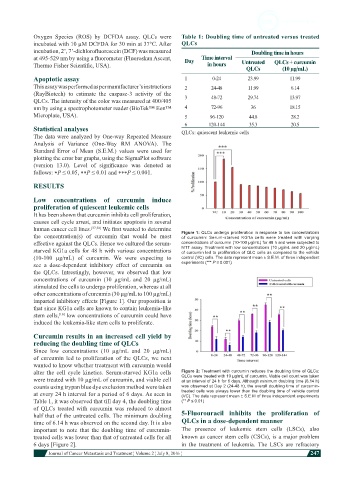
Oxygen Species (ROS) by DCFDA assay. QLCs were    Table 1: Doubling time of untreated versus treated
            incubated with 10 µM DCFDA for 30 min at 37°C. After   QLCs
            incubation, 2’, 7’-dichlorofluorescein (DCF) was measured                 Doubling time in hours
            at 495-529 nm by using a fluorometer (Fluoroskan Ascent,   Day  Time interval
            Thermo Fisher Scientific, USA).                            in hours    Untreated   QLCs + curcumin
                                                                                    QLCs        (10 µg/mL)
            Apoptotic assay                                    1         0-24        23.99         11.99
            This assay was performed as per manufacturer’s instructions   2  24-48   11.99         6.14
            (RayBiotech)  to estimate  the caspase-3 activity  of the   3  48-72     29.74         13.97
            QLCs. The intensity of the color was measured at 400/405
            nm by using a spectrophotometer reader (BioTek™ Eon™   4    72-96         36           18.15
            Microplate, USA).                                  5        96-120       44.8          28.2
                                                               6       120-144       35.3          20.5
            Statistical analyses                              QLCs: quiescent leukemic cells
            The data were analyzed by One-way Repeated Measure
            Analysis  of  Variance  (One-Way  RM  ANOVA).  The
            Standard  Error  of Mean  (S.E.M.) values  were  used for
            plotting the error bar graphs, using the SigmaPlot software
            (version  13.0).  Level  of  significance  was  denoted  as
            follows: ∗P ≤ 0.05, ∗∗P ≤ 0.01 and ∗∗∗P ≤ 0.001.

            RESULTS

            Low concentrations of curcumin induce
            proliferation of quiescent leukemic cells
            It has been shown that curcumin inhibits cell proliferation,
            causes cell cycle arrest, and initiates apoptosis in several
            human cancer cell lines. [37,38]  We first wanted to determine   Figure 1: QLCs undergo proliferation in response to low concentrations
            the concentration(s)  of curcumin that would be most   of curcumin: Serum-starved KG1a cells were treated with varying
            effective against the QLCs. Hence we cultured the serum-  concentrations of curcumin (10-100 µg/mL) for 48 h and were subjected to
            starved KG1a cells for 48 h with various concentrations   MTT assay. Treatment with low concentrations (10 µg/mL and 20 µg/mL)
                                                              of curcumin led to proliferation of QLC cells as compared to the vehicle
            (10-100  µg/mL)  of curcumin.  We  were  expecting  to   control (VC) cells. The data represent mean ± S.E.M. of three independent
            see a dose-dependent inhibitory effect  of curcumin  on   experiments (*** P ≤ 0.001)
            the QLCs.  Intrestingly, however, we observed that low
            concentrations  of curcumin  (10 µg/mL and  20 µg/mL)
            stimulated the cells to undergo proliferation, whereas at all
            other concentrations of curcumin (30 µg/mL to 100 µg/mL)
            imparted inhibitory effects [Figure 1]. Our proposition is
            that since KG1a cells are known to contain leukemia-like
            stem cells,  low concentrations of curcumin could have
                     [16]
            induced the leukemia-like stem cells to proliferate.
            Curcumin results in an increased cell yield by
            reducing the doubling time of QLCs
            Since low concentrations (10 µg/mL and 20 µg/mL)
            of curcumin led to proliferation of the QLCs, we next
            wanted to know whether treatment with curcumin would
            alter  the cell cycle  kinetics. Serum-starved  KG1a cells   Figure 2: Treatment with curcumin reduces the doubling time of QLCs:
            were treated with 10 µg/mL of curcumin, and viable cell   QLCs were treated with 10 µg/mL of curcumin. Viable cell count was taken
                                                              at an interval of 24 h for 6 days. Although minimum doubling time (6.14 h)
            counts using trypan blue dye exclusion method were taken   was observed at Day 2 (24-48 h), the overall doubling time of curcumin-
            at every 24 h interval for a period of 6 days. As seen in   treated cells was always lower than the doubling time of vehicle control
                                                              (VC). The data represent mean ± S.E.M of three independent experiments
            Table 1, it was observed that till day 4, the doubling time   (** P ≤ 0.01)
            of QLCs treated with curcumin was reduced to almost
            half that of the untreated cells. The minimum doubling   5-Fluorouracil inhibits the proliferation of
            time of 6.14 h was observed on the second day. It is also   QLCs in a dose-dependent manner
            important  to  note  that  the  doubling  time  of  curcumin-  The  presence  of  leukemic  stem  cells  (LSCs),  also
            treated cells was lower than that of untreated cells for all   known as cancer stem cells (CSCs), is a major problem
            6 days [Figure 2].                                in the treatment of leukemia.  The LSCs are refractory
                         Journal of Cancer Metastasis and Treatment ¦ Volume 2 ¦ July 8, 2016 ¦           247