
Page 24 - Read Online
P. 24
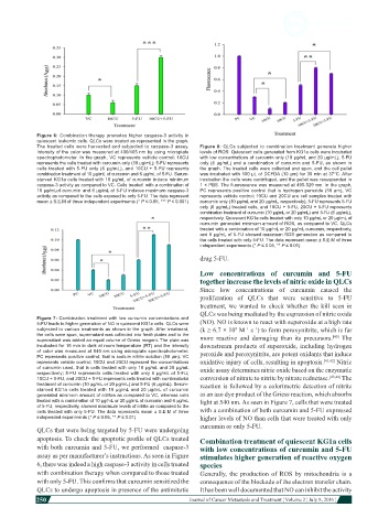
Figure 6: Combination therapy promotes higher caspase-3 activity in
            quiescent leukemic cells. QLCs were treated as represented in the graph.
            The treated cells were harvested and subjected to caspase-3 assay.   Figure 8: QLCs subjected to combination treatment generate higher
            Intensity of the color was measured at 400/405 nm by using microplate   levels of ROS: Quiescent cells generated from KG1a cells were incubated
            spectrophotometer. In the graph, VC represents vehicle control; 10CU   with low concentrations of curcumin only (10 µg/mL and 20 µg/mL), 5-FU
            represents the cells treated with curcumin only (10 µg/mL); 5-FU represents   only (6 µg/mL) and a combination of curcumin and 5-FU, as shown in
            cells treated with 5-FU only (6 µg/mL), and 10CU + 5-FU represents   the graph. The treated cells were collected and spun, and the cell pellet
            combination treatment of 10 µg/mL of curcumin and 6 µg/mL of 5-FU. Serum-  was incubated with 100 µL of DCFDA (10 µm) for 30 min at 37°C. After
            starved KG1a cells treated with 10 µg/mL of curcumin induce minimum   incubation the cells were centrifuged, and the pellet was resuspended in
            caspase-3 activity as compared to VC. Cells treated  with a combination of   1 × PBS. The fluorescence was measured at 495-529 nm. In the graph,
            10 µg/mLof curcumin and 6 µg/mL of 5-FU induces maximum caspase-3   PC represents positive control that is hydrogen peroxide (50 µm), VC
            activity as compared to the cells exposed to only 5-FU. The data represent   represents vehicle control; 10CU and 20CU are cell samples treated with
            mean ± S.E.M of three independent experiments (* P ≤ 0.05, *** P ≤ 0.001)  curcumin only (10 µg/mL and 20 µg/mL, respectively), 5-FU represents 5-FU
                                                              only (6 µg/mL) treated cells, and 10CU + 5-FU, 20CU + 5-FU represents
                                                              combination treatment of curcumin (10 µg/mL or 20 µg/mL) and 5-FU (6 µg/mL),
                                                              respectively. Quiescent KG1a cells treated with only 10 µg/mL or 20 µg/mL of
                                                              curcumin generated minimum amount of ROS, as compared to VC. QLCs
                                                              treated with a combination of 10 µg/mL or 20 µg/mL curcumin, respectively,
                                                              and 6 µg/mL of 5-FU showed maximum ROS generation as compared to
                                                              the cells treated with only 5-FU. The data represent mean ± S.E.M of three
                                                              independent experiments (* P ≤ 0.05, ** P ≤ 0.01)
                                                              drug 5-FU.

                                                              Low concentrations of curcumin and 5-FU
                                                              together increase the levels of nitric oxide in QLCs
                                                              Since  low concentrations  of curcumin  caused  the
                                                              proliferation  of QLCs that  were sensitive  to 5-FU
                                                              treatment,  we wanted  to check  whether  the  kill  seen in
                                                              QLCs was being mediated by the expression of nitric oxide
            Figure 7: Combination treatment with low curcumin concentrations and
            5-FU leads to higher generation of NO in quiescent KG1a cells: QLCs were   (NO). NO is known to react with superoxide at a high rate
                                                                         9
            subjected to various treatments as shown in the graph. After treatment,   (k ≥ 6.7 × 10  M  s ) to form peroxynitrite, which is far
                                                                               −1
                                                                            −1
            the cells were spun, supernatant was collected into fresh plates and to the   more  reactive  and damaging  than  its  precursors.   The
                                                                                                       [40]
            supernatant was added an equal volume of Griess reagent. The plate was
            incubated for 15 min in dark at room temperature (RT) and the intensity   downstream products of superoxide, including hydrogen
            of color was measured at 540 nm using microplate spectrophotometer.   peroxide and peroxynitrite, are potent oxidants that induce
            PC represents positive control, that is sodium nitrite solution (50 µm). VC
            represents vehicle control; 10CU and 20CU represent the concentrations   oxidative injury of cells, resulting in apoptosis. [41,42]  Nitric
            of curcumin used, that is cells treated with only 10 µg/mL and 20 µg/mL   oxide assay determines nitric oxide based on the enzymatic
            respectively; 5-FU represents cells treated with only 6 µg/mL of 5-FU;
            10CU + 5-FU, and 20CU + 5-FU represents cells treated with combinatorial   conversion of nitrate to nitrite by nitrate reductase. [43,44]  The
            treatment of curcumin (10 µg/mL or 20 µg/mL) and 5-FU (6 µg/mL). Serum-  reaction is followed by a colorimetric detection of nitrite
            starved KG1a cells treated with 10 µg/mL and 20 µg/mL of curcumin
            generated minimum amount of nitrites as compared to VC, whereas cells   as an azo dye product of the Griess reaction, which absorbs
            treated with a combination of 10 µg/mL or 20 µg/mL of curcumin and 6 µg/mL   light at 540 nm. As seen in Figure 7, cells that were treated
            of 5-FU, respectively, showed maximum levels of nitrite as compared to the
            cells treated with only 5-FU. The data represents mean ± S.E.M of three   with a combination of both curcumin and 5-FU expressed
            independed experiments (* P ≤ 0.05, ** P ≤ 0.01)  higher levels of NO than cells that were treated with only
                                                              curcumin or only 5-FU.
            QLCs that were being targeted by 5-FU were undergoing
            apoptosis. To check the apoptotic profile of QLCs treated   Combination treatment of quiescent KG1a cells
            with both curcumin and 5-FU, we performed  caspase-3   with low concentrations of curcumin and 5-FU
            assay as per manufacturer’s instructions. As seen in Figure   stimulates higher generation of reactive oxygen
            6, there was indeed a high caspase-3 activity in cells treated   species
            with combination therapy when compared to those treated   Generally, the  production  of ROS by mitochondria  is a
            with only 5-FU. This confirms that curcumin sensitized the   consequence of the blockade of the electron transfer chain.
            QLCs to undergo apoptosis in presence of the antimitotic   It has been well documented that NO can inhibit the activity
            250
                                                                                                                          Journal of Cancer Metastasis and Treatment ¦ Volume 2 ¦ July 8, 2016 ¦