
Page 33 - Read Online
P. 33
only the equipment, but also revision of the local of
implementation, making it a high cost technology. [40,41]
In addition, the time for image acquiring and the need
of stop the surgery for it, prolong time of surgery and
anesthesia. [42-44]
Roder et al. studied retrospectively 117 patients after
conventional surgery, after 5-ALA, and after iMRI they
found that mean residual tumor volume after iMRI-assisted
surgery (0.5 [0.0e4.7] cm ) was significantly smaller
3
compared to the residual tumor volume after 5-ALA-
guided surgery (1.9 [0.0-13.2] cm ; P = 0.022), which
3
was significantly smaller than in conventional surgery
(4.7 [0.0-30.6] cm ; P = 0.007). Total resections were
3
significantly more common in iMRI (74%) than in 5-ALA-
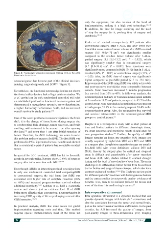
Figure 3: Tractography magnetic resonance imaging. In blue: the white assisted (46%, P = 0.05) or conventional surgery (13%, P
fibers tracts. In red: tumor = 0.03). Also, the iMRI time of surgery was significantly
neuronavigation has become part of the clinical decision- higher compared to pre-iMRI period (213 vs. 354 min).
making, surgical approach, and EOR [Figure 3]. Improvement of the EOR using iMRI was safely achievable
[33]
and post-operative morbidities were comparable between
Nevertheless, the functional neuronavigation has not shown cohorts. Total resections increased 6 months progression
its clinical utility due to a lack of high evidence studies. Wu free survival from 32% to 45%. In follow-up analysis, the
et al. carried out the only randomized controlled trial with neuronavigation had new or worsened neurological deficits
an established protocol in functional neuronavigation and at 3 months in 18.2% of patients, compared to 45.5% of the
demonstrated a reduced post-operative motor deterioration, control group. Non-neurological complications were present
a higher Karnofsky Performance Scale, and an increased in both groups, 31.8% in the control group and 30.4% in the
overall survival in study patients. [33] neuronavigation group. Also, the progression-free survival
and survival ratewasbetter in the neuronavigation/iMRI
One of the worst problems in neuronavigation is the brain groups vs. control groups. [2]
shift; it is the change of tissue/lesion during surgery due
to cerebrospinal fluid drainage, tumor resection, and brain Despite it is a retrospective study with a short period of
swelling; with estimated to be around 1 cm after opening time and limited patients in different chronologic times,
the dura, and more than 1 cm after initial resection of the great outcomes and promising results should open for
[34]
[42]
tumor. Therefore, the iMRI technology has come to solve new prospective studies. Further, the quality of iMRI
this problem and also increase the EOR. The first iMRI was images remains an issue; pre-operative MRI images are
performed in 1994; it presented several benefits and showed usually acquired by high-fields MRI with DTI and fMRI
that a considerable part of patients had resectable residual as a surgery plan, though intra-operative images are usually
tumor. low-field MRI with worse definitions without DTI and
fMRI; thereby the surgery plan for critical and eloquent
In special for LGG treatment, iMRI has led to favorable areas is difficult and questionable after tumor resection
results in several studies. Reports show 30-60% of return to and brain shift. Also, studies related to contrast dosage/
surgery after initial resection with iMRI. [35-38] timing and the local of resection have been done. The main
challenge is to differentiate tumor border from blood brain
Even though iMRI is an interesting method, nowadays there barrier brakes and surgical tissue changes, which also have
is only one randomized controlled trial comparingiMRI contrast-enchanted borders. [43,44] The Cochrane review point
to conventional surgery; the trial found that iMRI was for different patients’ baselines with heterogeneous lesions
associated with higher rate of complete resection (96% and the current studies do not provide quality evidences of
vs. 68%) and increased progression-free survival without benefits. Also, there is no standard protocol for its use and
additional morbidity. [5,39] Kubben et al. held a systematic most of the time it is used in single centers. [2]
review and showed just an evidence level II of iMRI
being more effective than conventional neuronavigation in Intra-operative ultrasound
increasing EOR, quality of life or prolonging survival after Intra-operative ultrasound is a dynamic method that can
GBM resection. [38,39] provide dynamic images with brain shift corrections and
also the correlation between the tumor and normal brain,
In practical analysis, iMRI has some issues for global just as the tumor vascular nutrition and borders. In the past
implementation regarding costs and time. This method decades, the iUS increased the quality of images, from
requires special implementation; most of the times not poor-quality images to three-dimensional (3D) imaging
74
Journal of Cancer Metastasis and Treatment ¦ Volume 2 ¦ March 11, 2016 ¦