
Page 31 - Read Online
P. 31
of improving survival rate. [2] hemisphere and localization of speech areas. The pre-motor
areas of the face are always tested to identify possible
In this point we have two arms, the technologies to improve motor causes of the aphasia. Even with all protocols,
resection and to increase the knowledge of tumor nature. By intra-operative positive sites errors can range from 4.6%
now it is clear that just improving resection won’t provide to 22%. The counting test is used to document a speech
[16]
the best result, but better understanding of the different arrest during electrocortical stimulation and also auditory
diseases and tumor natures, will provide direction for naming, verb generation and reading are commonly used
optimal resections and better outcomes. tests. Additional tests can be applied such as calculation,
visuospatial functions, working memory, visual pathways,
Awake craniotomy eye movements, and writing. [16,17]
Anesthetic advances permitted safer awake craniotomies
to obtain brain mapping and better neurosurgical borders. One important point is that function can be found at the
However, it has a series of challenges to be analyzed edge of high-grade gliomas and also within the tumor in
before such as integration of different types of knowledge, low-grades, so it has to be analyzed for a safe EOR. [19,20]
imaging, multidisciplinary team, cooperation from several
clinics sectors, application of protocols, application of Awake surgery has been used for some time, but new
specific technical adjustments, and a multidisciplinary tests and anesthetic evolution have permitted a better
approach. The integration of the pre-operative functional understanding of functional areas and also the mapping of
MRI (fMRI) and neuropsychological tests are the key for a complex brain areas.
good planning and patient selection. Not all tumor patients
should undergo awake craniotomy, but patients with lesions Cortical and sub-cortical mapping
close relationship with eloquent areas, in special for motor During the past years, the increase importance of the EOR
and speech. [16,17] Talacchi et al. stated that intra-operative and the relationship with increased overall survivalhas
complication can vary from anesthetic (inadequate or made the neurosurgeons push to the limits of the glioma
excessive sedation, pain, nausea, vomiting); respiratory surgeries, even in eloquent areas. Nevertheless, without
(oxygen saturation < 90%, increased CO , hypoventilation intra-operative monitoring, morbidity increasing became
2
< 8 breaths/min, airway obstruction); hemodynamic (hyper- fact. The objective of increasing overall survival with good
[1,21]
or hypotension, tachy- or bradycardia); and neurological functional status made the neuronavigation era a reality.
(convulsions, brain swelling, new neurological deficit). As imaging has increased its accuracy over the past years,
From these complications, hyper- and hypotension are neuroanatomy studies have shown a better knowledge of
the most frequent in awake surgery (11% and 56%, the sub-cortical tracts and the new mapping technologies
respectively). [16,17] have shown the real cortical and functional mapping, which
most of the times can be changed by the lesion. [9,22]
The main purpose of awake surgery is the monitoring of
speech and motor pathways. This way, the physical pre- Intra-operative monitoring has been studied by several
operative imaging/clinical examinations and intra-operative different methods, using somatosensory-evoked potentials
positive tests are important. Patients with aphasias and (SSEP), awake stimulation, and cortical/sub-cortical direct
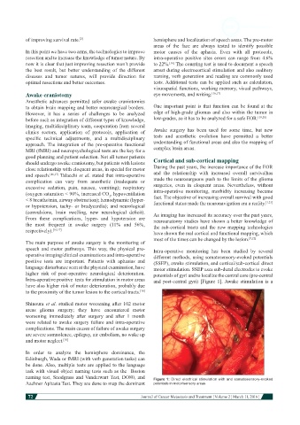
language disturbance seen at the physical examination, have motor stimulation. SSEP uses sub-dural electrodes to evoke
higher risk of post-operative neurological deterioration. potentials of gyri and to localize the central core (pre-central
Intra-operative positive tests for stimulation in motor areas and post-central gyri) [Figure 1]. Awake stimulation is a
have also higher risk of motor deterioration, probably due
to the proximity of the tumor lesion to the cortical tracts. [16]
Shinoura et al. studied motor worsening after 102 motor
areas glioma surgery; they have encountered motor
worsening immediately after surgery and after 1 month
were related to awake surgery failure and intra-operative
complications. The main causes of failure of awake surgery
are severe somnolence, epilepsy, air embolism, no wake up
and motor neglect. [18]
In order to analyze the hemisphere dominance, the
Edinburgh, Wada or fMRI (with verb generation tasks) can
be done. Also, multiple tests are applied to the language
task with visual object naming tests such as the Boston
naming test, Snodgrass and Vanderwart Test, DO80, and Figure 1: Direct electrical stimulation with and somatosensory-evoked
Aachner Aphasia Test. They are done to map the dominant potentials in motor/sensory areas
72
Journal of Cancer Metastasis and Treatment ¦ Volume 2 ¦ March 11, 2016 ¦