
Page 59 - Read Online
P. 59
Farkas et al. J Cancer Metastasis Treat 2022;8:37 https://dx.doi.org/10.20517/2394-4722.2022.89 Page 5 of 13
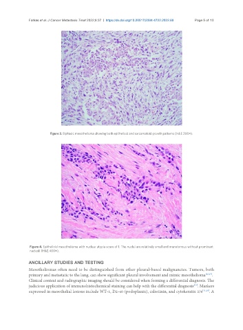
Figure 3. Biphasic mesothelioma showing both epithelioid and sarcomatoid growth patterns (H&E 200×).
Figure 4. Epithelioid mesothelioma with nuclear atypia score of 1. The nuclei are relatively small and monotonous without prominent
nucleoli (H&E 400×).
ANCILLARY STUDIES AND TESTING
Mesotheliomas often need to be distinguished from other pleural-based malignancies. Tumors, both
primary and metastatic to the lung, can show significant pleural involvement and mimic mesothelioma [26,27] .
Clinical context and radiographic imaging should be considered when forming a differential diagnosis. The
[11]
judicious application of immunohistochemical staining can help with the differential diagnosis . Markers
expressed in mesothelial lesions include WT-1, D2-40 (podoplanin), calretinin, and cytokeratin 5/6 [11,28] . A