
Page 60 - Read Online
P. 60
Page 6 of 13 Farkas et al. J Cancer Metastasis Treat 2022;8:37 https://dx.doi.org/10.20517/2394-4722.2022.89
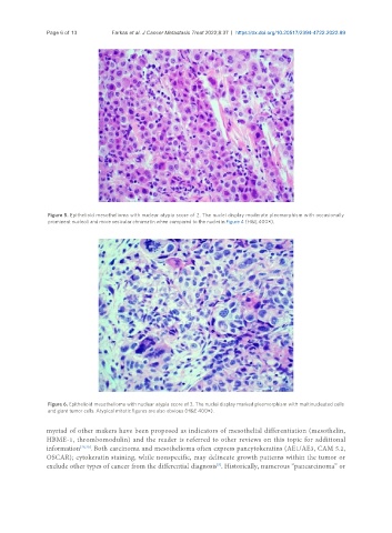
Figure 5. Epithelioid mesothelioma with nuclear atypia score of 2. The nuclei display moderate pleomorphism with occasionally
prominent nucleoli and more vesicular chromatin when compared to the nuclei in Figure 4 (H&E 400×).
Figure 6. Epithelioid mesothelioma with nuclear atypia score of 3. The nuclei display marked pleomorphism with multinucleated cells
and giant tumor cells. Atypical mitotic figures are also obvious (H&E 400×).
myriad of other makers have been proposed as indicators of mesothelial differentiation (mesothelin,
HBME-1, thrombomodulin) and the reader is referred to other reviews on this topic for additional
information [29,30] . Both carcinoma and mesothelioma often express pancytokeratins (AE1/AE3, CAM 5.2,
OSCAR); cytokeratin staining, while nonspecific, may delineate growth patterns within the tumor or
[5]
exclude other types of cancer from the differential diagnosis . Historically, numerous “pancarcinoma” or