
Page 45 - Read Online
P. 45
Galli et al. J Cancer Metastasis Treat 2022;8:48 https://dx.doi.org/10.20517/2394-4722.2022.19 Page 7 of 14
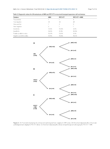
Table 3. Diagnostic values for LN metastases of MRI and PET/CT in recurrent laryngeal squamous cell carcinomas
Feature MRI PET/CT PET/CT + MRI
True positive 1 1 0
True negative 31 30 25
False positive 1 3 3
False negative 2 1 1
Sensitivity 33.3% 50.0% 0.0%
Specificity 96.9% 91.0% 89.3%
Positive predictive value 50.0% 25.0% 0.0%
Negative predictive value 93.9% 96.8% 96.2%
Figure 2. (A) Flowchart displaying the clinical and pathological neck staging for MRI alone. (B) flowchart displaying the clinical and
pathological neck staging for PET/CT alone. (C) flowchart displaying the clinical and pathological neck staging for PET/CT + MRI.