
Page 95 - Read Online
P. 95
Page 14 of 20 Ottewell et al. J Cancer Metastasis Treat 2021;7:11 https://dx.doi.org/10.20517/2394-4722.2021.14
5
5
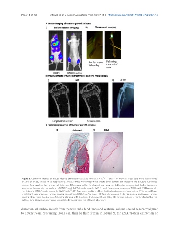
Figure 3. Common analysis of mouse models of bone metastases. In total, 1 × 10 4T1 or 5 × 10 MDA-MB-231 cells were injected into
BALB/c or BALB/c nude mice, respectively. BALB/c mice were imaged two weeks after tumour cell injection and BALB/c nude mice
imaged four weeks after tumour cell injection. Mice were culled for downstream analysis 24 h after imaging. (A) Bioluminescence
imaging of tumours in the skeleton of BALB/c and BALB/c nude mice by IVIS (I) and florescence imaging of MDA-MB-231 tumours in
TM
the tibia of a BALB/c nude mouse by LightTools . (B) Two-cross sections of longitudinal and cross-sectional micro-CT images (I) and
whole leg X-ray images of tumour bearing bones from BALB/c nu/nu mice. (C) Two-dimensional 3 µM histological sections of tumour
bearing tibiae from BALB/c mice following staining with Goldner’s trichrome (I) and H&E (II); tumour in bone is highlighted with a red
outline. Data shown are previously unpublished images from the Ottewell laboratory.
dissection, all skeletal muscle from the forelimbs, hind limbs and vertebral column should be removed prior
to downstream processing. Bone can then be flash frozen in liquid N for RNA/protein extraction or
2