
Page 91 - Read Online
P. 91
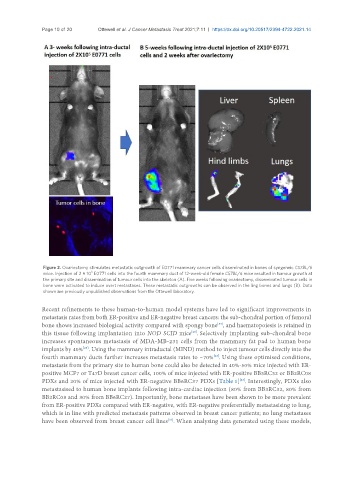
Page 10 of 20      Ottewell et al. J Cancer Metastasis Treat 2021;7:11  https://dx.doi.org/10.20517/2394-4722.2021.14


                Figure 2. Ovariectomy stimulates metastatic outgrowth of E0771 mammary cancer cells disseminated in bones of syngeneic C57BL/6
                                5
                mice. Injection of 2 × 10  E0771 cells into the fourth mammary duct of 12-week-old female C57BL/6 mice resulted in tumour growth at
                the primary site and dissemination of tumour cells into the skeleton (A). Five weeks following ovariectomy, disseminated tumour cells in
                bone were activated to induce overt metastases. These metastatic outgrowths can be observed in the ling bones and lungs (B). Data
                shown are previously unpublished observations from the Ottewell laboratory.

               Recent refinements to these human-to-human model systems have led to significant improvements in
               metastasis rates from both ER-positive and ER-negative breast cancers: the sub-chondral portion of femoral
                                                                          [53]
               bone shows increased biological activity compared with spongy bone , and haematopoiesis is retained in
               this tissue following implantation into NOD SCID mice . Selectively implanting sub-chondral bone
                                                                 [23]
               increases spontaneous metastasis of MDA-MB-231 cells from the mammary fat pad to human bone
               implants by 40% . Using the mammary intraductal (MIND) method to inject tumour cells directly into the
                             [23]
               fourth mammary ducts further increases metastasis rates to ~70% . Using these optimised conditions,
                                                                         [23]
               metastasis from the primary site to human bone could also be detected in 40%-50% mice injected with ER-
               positive MCF7 or T47D breast cancer cells, 100% of mice injected with ER-positive BB3RC32 or BB2RC08
               PDXs and 20% of mice injected with ER-negative BB6RC37 PDXs [Table 1] . Interestingly, PDXs also
                                                                                  [23]
               metastasised to human bone implants following intra-cardiac injection (80% from BB3RC32, 80% from
               BB2RC08 and 30% from BB6RC27). Importantly, bone metastases have been shown to be more prevalent
               from ER-positive PDXs compared with ER-negative, with ER-negative preferentially metastasising to lung,
               which is in line with predicted metastasis patterns observed in breast cancer patients; no lung metastases
                                                          [23]
               have been observed from breast cancer cell lines . When analysing data generated using these models,