
Page 97 - Read Online
P. 97
Page 16 of 20 Ottewell et al. J Cancer Metastasis Treat 2021;7:11 https://dx.doi.org/10.20517/2394-4722.2021.14
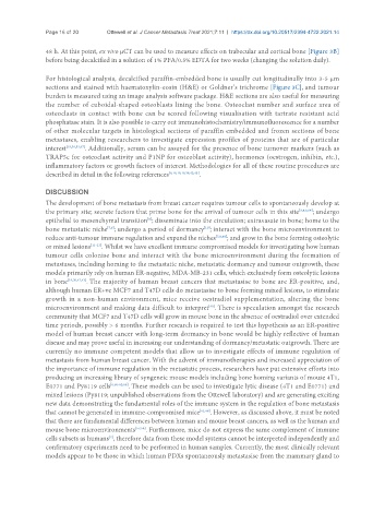
48 h. At this point, ex vivo µCT can be used to measure effects on trabecular and cortical bone [Figure 3B]
before being decalcified in a solution of 1% PFA/0.5% EDTA for two weeks (changing the solution daily).
For histological analysis, decalcified paraffin-embedded bone is usually cut longitudinally into 3-5 µm
sections and stained with haematoxylin-eosin (H&E) or Goldner’s trichrome [Figure 3C], and tumour
burden is measured using an image analysis software package. H&E sections are also useful for measuring
the number of cuboidal-shaped osteoblasts lining the bone. Osteoclast number and surface area of
osteoclasts in contact with bone can be scored following visualisation with tartrate resistant acid
phosphatase stain. It is also possible to carry out immunohistochemistry/immunofluorescence for a number
of other molecular targets in histological sections of paraffin embedded and frozen sections of bone
metastases, enabling researchers to investigate expression profiles of proteins that are of particular
interest [19,30,53,67] . Additionally, serum can be assayed for the presence of bone turnover markers (such as
TRAP5c for osteoclast activity and P1NP for osteoblast activity), hormones (oestrogen, inhibin, etc.),
inflammatory factors or growth factors of interest. Methodologies for all of these routine procedures are
described in detail in the following references [6,16,18,19,36,42,43] .
DISCUSSION
The development of bone metastasis from breast cancer requires tumour cells to spontaneously develop at
the primary site; secrete factors that prime bone for the arrival of tumour cells in this site [3,4,6,64] ; undergo
epithelial to mesenchymal transition ; disseminate into the circulation; extravasate in bone; home to the
[6]
[10]
bone metastatic niche ; undergo a period of dormancy ; interact with the bone microenvironment to
[7,8]
reduce anti-tumour immune regulation and expand the niches [7,8,68] ; and grow in the bone forming osteolytic
or mixed lesions [11-13] . Whilst we have excellent immune compromised models for investigating how human
tumour cells colonise bone and interact with the bone microenvironment during the formation of
metastases, including homing to the metastatic niche, metastatic dormancy and tumour outgrowth, these
models primarily rely on human ER-negative, MDA-MB-231 cells, which exclusively form osteolytic lesions
in bone [19,30,42,43] . The majority of human breast cancers that metastasise to bone are ER-positive, and,
although human ER+ve MCF7 and T47D cells do metastasise to bone forming mixed lesions, to stimulate
growth in a non-human environment, mice receive oestradiol supplementation, altering the bone
[40]
microenvironment and making data difficult to interpret . There is speculation amongst the research
community that MCF7 and T47D cells will grow in mouse bone in the absence of oestradiol over extended
time periods, possibly > 6 months. Further research is required to test this hypothesis as an ER-positive
model of human breast cancer with long-term dormancy in bone would be highly reflective of human
disease and may prove useful in increasing our understanding of dormancy/metastatic outgrowth. There are
currently no immune competent models that allow us to investigate effects of immune regulation of
metastasis from human breast cancer. With the advent of immunotherapies and increased appreciation of
the importance of immune regulation in the metastatic process, researchers have put extensive efforts into
producing an increasing library of syngeneic mouse models including bone homing variants of mouse 4T1,
E0771 and Py8119 cells [6,60-62,68] . These models can be used to investigate lytic disease (4T1 and E0771) and
mixed lesions (Py8119; unpublished observations from the Ottewell laboratory) and are generating exciting
new data demonstrating the fundamental roles of the immune system in the regulation of bone metastasis
that cannot be generated in immune-compromised mice [61,68] . However, as discussed above, it must be noted
that there are fundamental differences between human and mouse breast cancers, as well as the human and
mouse bone microenvironments [57,58] . Furthermore, mice do not express the same complement of immune
cells subsets as humans , therefore data from these model systems cannot be interpreted independently and
[2]
confirmatory experiments need to be performed in human samples. Currently, the most clinically relevant
models appear to be those in which human PDXs spontaneously metastasise from the mammary gland to