
Page 37 - Read Online
P. 37
Page 10 of 15 Pacheco et al. J Cancer Metastasis Treat 2020;6:49 I http://dx.doi.org/10.20517/2394-4722.2020.85
C
A B D
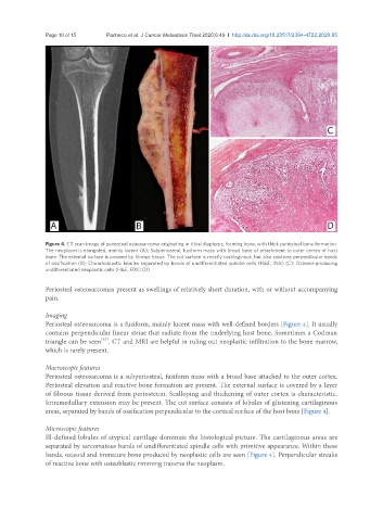
Figure 4. CT scan image of periosteal osteosarcoma originating in tibial diaphysis, forming bone, with thick periosteal bone formation.
The neoplasm is elongated, mainly lucent (A); Subperiosteal, fusiform mass with broad base of attachment to outer cortex of host
bone. The external surface is covered by fibrous tissue. The cut surface is mostly cartilaginous, but also contains perpendicular bands
of ossification (B); Chondroblastic lobules separated by bands of undifferentiated spindle cells (H&E; 25X) (C); Osteoid-producing
undifferentiated neoplastic cells (H&E, 50X) (D)
Periosteal osteosarcomas present as swellings of relatively short duration, with or without accompanying
pain.
Imaging
Periosteal osteosarcoma is a fusiform, mainly lucent mass with well-defined borders [Figure 4]. It usually
contains perpendicular linear striae that radiate from the underlying host bone. Sometimes a Codman
[1,3]
triangle can be seen . CT and MRI are helpful in ruling out neoplastic infiltration to the bone marrow,
which is rarely present.
Macroscopic features
Periosteal osteosarcoma is a subperiosteal, fusiform mass with a broad base attached to the outer cortex.
Periosteal elevation and reactive bone formation are present. The external surface is covered by a layer
of fibrous tissue derived from periosteum. Scalloping and thickening of outer cortex is characteristic.
Intramedullary extension may be present. The cut surface consists of lobules of glistening cartilaginous
areas, separated by bands of ossification perpendicular to the cortical surface of the host bone [Figure 4].
Microscopic features
Ill-defined lobules of atypical cartilage dominate the histological picture. The cartilaginous areas are
separated by sarcomatous bands of undifferentiated spindle cells with primitive appearance. Within these
bands, osteoid and immature bone produced by neoplastic cells are seen [Figure 4]. Perpendicular streaks
of reactive bone with osteoblastic rimming traverse the neoplasm.