
Page 39 - Read Online
P. 39
Page 12 of 15 Pacheco et al. J Cancer Metastasis Treat 2020;6:49 I http://dx.doi.org/10.20517/2394-4722.2020.85
A B
C D
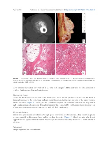
Figure 5. CT scan shows a tumor that develops in the soft tissue but starts from the cortex (A); High-grade surface osteosarcoma of
distal femur with cortical erosion (B); Infiltrating neoplasm in the surrounding soft tissues (H&E;25X) (C); Highly atypical features and
osteoid production (H&E; 50X) (D)
[3]
show minimal medullary involvement in CT and MRI images . MRI facilitates the identification of
cartilage that is scattered throughout the mass.
Macroscopic features
Lobulated, relatively well-circumscribed, broad-base mass on the periosteal surface of the bone. It
frequently extends to the periosteum and can erode the cortex, but the vast majority of the tumor remains
outside the bone [Figure 5]. Any significant penetration beyond the endosteum exclude the diagnosis of
high- grade surface osteosarcoma. The cut surface may be dominated by cartilaginous tissue or composed
of hard, tan-white areas admixed with others with fish flesh consistency.
Microscopic features
The microscopic features are identical to high-grade conventional osteosarcoma. They exhibit anaplasia,
necrosis, osteoid, and immature bone and/or cartilage formation [Figure 5]. Mitotic activity is brisk, and
atypical mitotic figures are easily found. Microscopic evidence of medullary extension is either absent or
minimal.
Pathogenesis
The pathogenesis remains unknown.