
Page 32 - Read Online
P. 32
Pacheco et al. J Cancer Metastasis Treat 2020;6:49 I http://dx.doi.org/10.20517/2394-4722.2020.85 Page 5 of 15
A B
C D
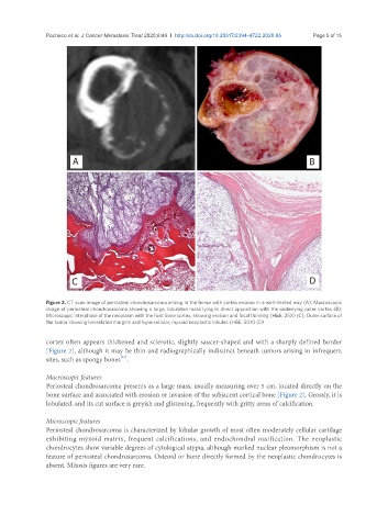
Figure 2. CT scan image of periosteal chondrosarcoma arising in the femur with cortex erosion in a well-limited way (A); Macroscopic
image of periosteal chondrosarcoma showing a large, lobulated mass lying in direct apposition with the underlying outer cortex (B);
Microscopic interphase of the neoplasm with the host bone cortex, showing erosion and focal thinning (H&E; 25X) (C); Outer surface of
the tumor showing bosselated margins and hypercellular, myxoid neoplastic lobules (H&E; 50X) (D)
cortex often appears thickened and sclerotic, slightly saucer-shaped and with a sharply defined border
[Figure 2], although it may be thin and radiographically indistinct beneath tumors arising in infrequent
sites, such as spongy bones .
[21]
Macroscopic features
Periosteal chondrosarcoma presents as a large mass, usually measuring over 5 cm, located directly on the
bone surface and associated with erosion or invasion of the subjacent cortical bone [Figure 2]. Grossly, it is
lobulated, and its cut surface is greyish and glistening, frequently with gritty areas of calcification.
Microscopic features
Periosteal chondrosarcoma is characterized by lobular growth of most often moderately cellular cartilage
exhibiting myxoid matrix, frequent calcifications, and endochondral ossification. The neoplastic
chondrocytes show variable degrees of cytological atypia, although marked nuclear pleomorphism is not a
feature of periosteal chondrosarcoma. Osteoid or bone directly formed by the neoplastic chondrocytes is
absent. Mitosis figures are very rare.