
Page 30 - Read Online
P. 30
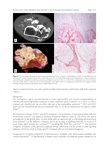
Pacheco et al. J Cancer Metastasis Treat 2020;6:49  I  http://dx.doi.org/10.20517/2394-4722.2020.85                    Page 3 of 15


                 A                                                                                     C


                 B                                                                                    D


               Figure 1. CT scan image of femoral peripheral secondary chondrosarcoma in patient with hereditary multiple osteochondromas with
               characteristic cortical flaring at the junction of the host bone with the stalk of the neoplasia (A); Macroscopic image of exophytic
               osteocartilaginous neoplasm containing a glistening cartilaginous cap more than 2 cm thick (B); Cartilaginous cap at the interphase
               with bone stalk, which is continuous with the host medullary canal (H&E; 25X) (C); Disorganized, hypercellular and atypical hyaline
               cartilage arranged in lobules separated by fibrous bands (H&E; 50X) (D)

               There is continuity between the cortex and the medulla of the host bone and the bony stalk of the neoplasm
               [Figure 1].

               Pathogenesis
               The cartilaginous cap in osteochondromas is mosaic (genetically), and contains a subpopulation of
               chondrocytes harboring biallelic mutations of tumor-suppressor genes Exostosin-1 or 2 (EXT1 or EXT2),
                                                                                        [5-7]
               admixed with chondrocytes that are either wild-type or have monoallelic mutations . EXT1 and EXT2
               genes encode transmembrane glycosyltransferases that act in the polymerization of heparan sulfate
                     [5]
               chains .
               The functional outcome of EXT1 and EXT2 mutations is the production of truncated proteins with loss
               of enzymatic activity  and impaired synthesis of heparan sulphate chains on cell surface and matrix
                                  [8]
               proteoglycans. In the growth plates, heparan sulfate plays an important role in establishing and maintaining
                            [6,9]
               tissue polarity , in the diffusion of ligands, and in the binding of signaling molecules to receptors of
               intracellular signal transduction pathways [6,9,10]  including Ihh, BMP, Wnt, and FGF [10,11] . The impaired
               elongation of heparan sulfate glycosaminoglycans disrupt cell polarity and the activity of these signaling
               pathways, which may result in ectopic growth of mutated cells and osteochondromagenesis.


               Progression to secondary peripheral chondrosarcoma, a neoplasia with chromosomal instability and
               complex karyotypes , is hypothesized to happen upon acquisition of additional genetic changes by the
                                [12]