
Page 29 - Read Online
P. 29
Page 2 of 15 Pacheco et al. J Cancer Metastasis Treat 2020;6:49 I http://dx.doi.org/10.20517/2394-4722.2020.85
main clinicopathologic and radiologic features of malignant tumors of bone surface, to summarize their
pathogenesis and to offer a useful framework for their differential diagnosis with emphasis on potential
diagnostic difficulties.
CHONDROGENIC TUMORS
Secondary peripheral chondrosarcoma
Definition and clinical features
Secondary peripheral chondrosarcomas are cartilaginous malignant neoplasms arising from the chondroid
cap of pre-existent osteochondromas. They are graded into grades 1 to 3, according to the World Health
Organization (WHO) histological grading system used for central chondrosarcomas. Grade 1 neoplasms
receive the name of secondary peripheral atypical cartilaginous tumor if they arise in the appendicular
[2]
skeleton, and secondary peripheral chondrosarcoma grade 1 if they arise in axial locations .
[1,3]
They constitute approximately 12%-18% of all chondrosarcomas , and occur after puberty, with a
[1,4]
peak incidence between 20 and 40 years. Males are affected more than females . Secondary peripheral
chondrosarcomas most commonly arise in the bones of the pelvis (40% of cases). Other common locations
[1]
are the proximal and distal femur (19.5%), scapula (10%), vertebral column (9%), and ribs (5%) .
Rapid growth or onset of pain on a pre-existing osteochondroma after puberty, should raise suspicion of
malignant transformation, which occurs in 1% of solitary osteochondroma and in up to 5% of patients
with hereditary multiple osteochondromas. Depending on the location of the neoplasm, neurologic (e.g.,
numbness, weakness, radiating pain, paraplegia), urinary, or colonic symptoms may be present.
Imaging
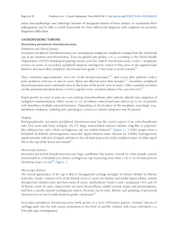
Radiographically, secondary peripheral chondrosarcoma has the typical aspect of an osteochondroma
with lytic areas and fuzzy margins. On CT, large, noncalcified tumoral lobules, ring-like or popcorn-
[1]
like radiopacities, and a thick cartilaginous cap are evident features [Figure 1]. T1MRI images show a
lobulated, ill-defined, inhomogeneous muscular signal intensity mass; whereas on T2MRI, heterogeneous
signal intensity with foci of signal void due to the calcified areas and a thick peripheral layer of white signal
[1]
due to the cap of the lesion are evident .
Macroscopic features
Secondary peripheral chondrosarcomas are large, cauliflower-like masses covered by a thin pseudo-capsule
and beneath it, a lobulated and chalky cartilaginous cap measuring more than 2 cm in its thickest portion
(thickness mean 3.9 cm) [Figure 1].
[4]
Microscopic features
The overall appearance of the cap is that of disorganized cartilage arranged in lobules divided by fibrous
trabecula. Grade 1 tumors (65% of the Rizzoli series of cases) are hyaline and mildly hypercellular, contain
disorganized chondrocytes, and have areas of coarse calcifications. Grade 2 and 3 neoplasms (30% and 5%
of Rizzoli series of cases, respectively) are more hypercellular, exhibit nuclear atypia and pleomorphism,
and have a mostly myxoid cartilaginous matrix. Necrosis can be seen. Mitosis and spindling of peripheral
chondrocytes are more easily found in grade 3 neoplasms .
[1]
Secondary peripheral chondrosarcoma rarely grows in a truly infiltrative pattern. Instead, lobules of
cartilage push into the soft tissues, sometimes in the form of satellite nodules. Soft tissue infiltration is a
bona fide sign of malignancy.